
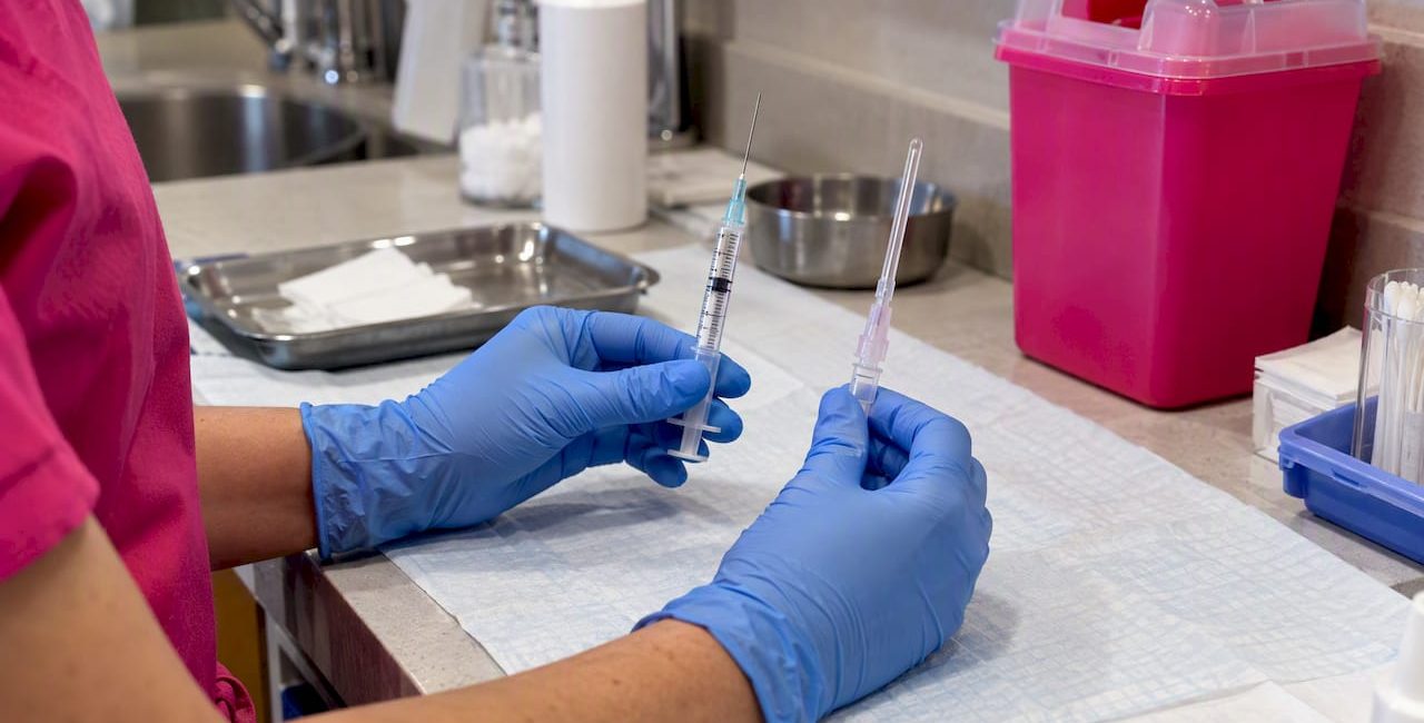
Cohort and review evidence points to cannulas carrying a lower signal for vascular occlusion than needles, though neither device is risk-free. In the largest reported dataset, needle injections carried an occlusion rate of roughly 1 in 6,410 compared with about 1 in 40,882 for cannulas, a 77.1% reduction in odds. Operator experience, injection plane and technique confound this figure, so the practical stance is to favour cannulas in vessel-dense, broad-distribution work while keeping needle skills sharp for structural and periosteal placement.
TL;DR:
- Cannulas significantly reduce vascular occlusion risk, with a 77.1% lower odds compared to needles in large studies, but they are not completely risk-free.
- Using a cannula in broad, vessel-dense areas minimizes trauma, while needles offer higher precision for structural or periosteal injections.
- Proper technique, including slow, low-pressure injections and ultrasound guidance, is crucial for safety regardless of device choice.
- Factors such as vessel depth, anatomical region, and product viscosity influence whether a needle or cannula is the better option for each specific task.
- Operator experience and site-specific anatomy remain key to reducing complications, emphasizing the need for skilled, individualized device selection.
Table of Contents
- Summary of the best evidence: cohorts, trials and reviews
- How cannulas and needles interact with vascular anatomy
- Device recommendations by anatomical region
- Injection technique and universal risk controls
- Situations where needles remain the better choice
- Mirror Pharma resources, author and practitioner tools
- A pragmatic view on adopting cannula technique
- Mirror Pharma: how we support safe practice for licensed clinicians
- FAQ
- Sources
Summary of the best evidence: cohorts, trials and reviews
The strongest data point comes from a 2020 retrospective cohort of 370 dermatologists reporting 1.7 million filler injections. Needle-based injections showed an occlusion rate of approximately 1 per 6,410 procedures, against roughly 1 per 40,882 for cannula-based injections.
Cannula use was linked to a 77.1% reduction in the odds of vascular occlusion compared with needles in the large retrospective cohort, making it one of the most frequently cited figures in this field.
Randomised evidence is thinner but consistent in direction. A double-blind trial comparing a metallic cannula with a standard needle for nasolabial fold augmentation randomised each side of the face within the same participants and found less pain, oedema and haematoma on the cannula-treated side by day three. The sample was small, and the finding is specific to that indication, but it supports the broader pattern seen in cohort data.
A 2025 narrative review synthesising randomised, observational and cadaveric evidence concluded that device choice should be individualised rather than fixed. Cannulas tend to reduce bruising and lower the vascular occlusion signal in vessel-dense regions, but needles remain preferable where precise periosteal or ligamentous placement is the clinical priority. The review’s authors argue against a dogmatic position: a blended approach using cannula for broad, low-pressure distribution and needle for precise structural support best reflects the current evidence base.
Several limitations run through this literature:
- Study endpoints vary widely, from angiographic occlusion to clinically reported bruising, which makes pooling results difficult.
- Operator experience is a major confounder that most observational studies cannot fully separate from device effect.
- Randomised trial volume remains small relative to the scale of real-world filler practice.
- Publication and reporting bias likely favour studies with a clear device-safety narrative over null results.
Taken together, the direction of effect is reasonably consistent: cannulas lower the measured rate of vascular occlusion in large samples, and reviews converge on individualised, region-specific use rather than a universal rule.
How cannulas and needles interact with vascular anatomy
The mechanical difference between the two devices explains much of the observed safety signal. A needle’s bevelled tip penetrates tissue and vessel walls directly along its line of travel. A cannula’s blunt tip tends to push vessels aside as it advances, displacing them laterally rather than piercing through, provided the tip meets the vessel wall at a shallow or glancing angle.
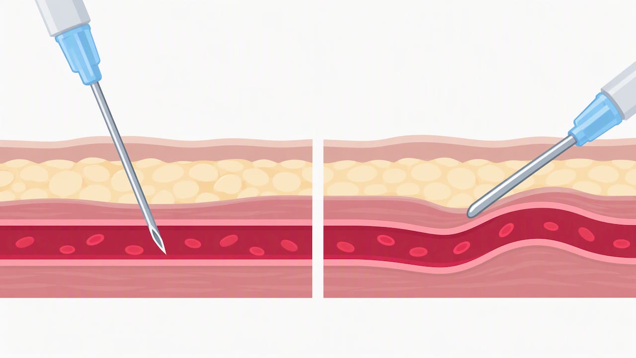
That protective effect is not absolute. Cadaveric evidence shows blunt cannulas usually reduce arterial perforation risk but can still perforate vessels under specific conditions, including perpendicular tip alignment, injection near vessel bifurcations, and high injection pressure forcing the tip through a vessel wall it would otherwise have displaced. A cadaveric systematic review of filler embolism mechanisms describes these failure modes in detail and uses them to underpin recommendations on injection angle and plane selection.
Several factors influence whether a cannula behaves safely in a given pass:
- Gauge and flexibility: a larger-gauge, more rigid cannula is less likely to flex around a vessel and more likely to displace or compress it rather than track alongside it.
- Tip shape and orientation: rounded, closed tips with side-ports generally reduce sharp contact compared with open-ended designs, though orientation relative to vessel course matters more than tip design alone.
- Angle of approach: a cannula advancing parallel to a vessel’s long axis is far less likely to penetrate it than one approaching perpendicular to the vessel wall.
- Injection pressure: high-pressure boluses can force fluid, and occasionally the cannula tip itself, through tissue planes that would otherwise resist penetration.
Pro Tip: Advance cannulas with a slow, deliberate fanning motion and keep the trajectory parallel to the expected vessel course rather than probing directly towards it.
Cadaveric studies offer excellent visibility of vessel architecture and failure mechanics, but they cannot reproduce live tissue turgor, blood flow or patient movement, so their angle and pressure thresholds should be read as guidance rather than fixed rules for every injector.
Device recommendations by anatomical region
Site-specific risk varies enormously across the face and body, and the literature supports different defaults depending on vessel density, depth and the structural demands of the area.
- Glabella: treat as a high-risk zone regardless of device; the supratrochlear and supraorbital vessels sit close to the surface here, and a cautious, low-volume approach with either device, informed by anatomical landmarking, is the conservative default.
- Nasal dorsum and tip: favour a supraperiosteal plane and midline trajectory where possible, since the nasal vasculature is variable and prone to serious occlusion; many practitioners use a cannula for broader dorsal work but revert to careful needle technique for very fine tip definition, always at low volume.
- Periorbital region: vessel density and the risk of retinal involvement make this an area for extreme caution with either device; small aliquots, slow injection and detailed anatomical knowledge matter more here than device choice alone.
- Cheek and midface: this is where cannulas generally show their clearest safety and comfort advantage, since broad subcutaneous fanning across a wider vessel-dense plane benefits from a blunt tip’s ability to glide past rather than through vessels.
- Hands: the dorsal hand has a dense, visible superficial venous network; a cannula’s lateral displacement effect suits the fanning technique commonly used here.
- Neck: similarly vessel-dense and best suited to broad, low-pressure distribution, which favours cannula technique over point-by-point needle injection.
- Lips and vermilion border: fine border definition and anchoring to the orbicularis oris often call for a needle’s precision, used with caution, small aliquots and aspiration, since this is a notoriously vascular area despite the relatively small vessels involved.
- Nasolabial folds and marionette lines: moderate-risk zones where the randomised trial evidence directly supports cannula use, given the documented reduction in bruising and discomfort in that specific comparison.
As a practical rule, gauge selection tends to run from around 25G to 27G for fine facial cannula work and up to 22G to 25G for broader body or deeper structural placement, with needle gauge chosen to match the viscosity of the product rather than to chase a safety effect on its own. Entry point and trajectory planning, favouring midline or parallel-to-vessel paths and a supraperiosteal plane in nasal work, matters at least as much as the device itself.
Injection technique and universal risk controls
Device choice sits inside a wider set of technique controls that apply whichever instrument is in hand. The 2026 systematic review of vascular occlusion management describes prevention as resting chiefly on anatomical planning, low-volume low-pressure injection and procedural standardisation, with cannula use and ultrasound treated as promising adjuncts rather than substitutes for sound technique.
- Inject in slow, retrograde micro-boluses using small aliquots, since this limits the volume delivered into any single tissue plane if a vessel is inadvertently compromised.
- Keep injection pressure low throughout; high-pressure boluses are a recognised contributor to vessel penetration and embolism risk with either device.
- Aspirate before injecting where the product and syringe allow, but treat a negative aspiration as reassurance rather than proof of a safe position, since false negatives occur with small-bore needles, viscous products and brief aspiration times.
- Reassess the treatment plan if the patient reports disproportionate pain, blanching or livedo patterning, since these are early signs that outrank any aspiration result.
Pro Tip: Treat aspiration as one layer of a safety system, not a stand-alone test; pair it with slow injection, small aliquots and constant visual and verbal monitoring of the patient.
Ultrasound guidance is increasingly recommended as a practical adjunct for high-risk areas and for practitioners adopting cannula techniques, since it allows direct visualisation of tissue plane and nearby vessels before and during injection. Its main limitations are the learning curve required to interpret real-time images accurately and the added procedure time, which means it supplements anatomical knowledge rather than replacing it.
Immediate management readiness is part of the same system. Expert protocol guidance recommends repeated, high-dose hyaluronidase infiltrated across the ischaemic territory and along the suspected vessel course as the central endpoint in managing a vascular occlusion, rather than relying on warm packs or massage as primary measures. Every clinic using dermal filler should have a written escalation pathway, a stocked hyaluronidase protocol, clear documentation standards and a plan for urgent referral where perfusion does not recover.
Situations where needles remain the better choice
Needles are not an inferior default; they are the right tool for specific jobs. Precise periosteal placement, where the product needs to sit directly against bone to support a structural correction, usually calls for a needle’s accuracy over a cannula’s blunt glide. Fine focal work using a true micro-bolus technique, where millimetre-level placement matters more than broad distribution, also tends to favour needles.
Patient history changes the calculation too. Prior surgery, scarring or distorted anatomy can make a cannula harder to pass cleanly through tissue planes, sometimes increasing the force needed and, paradoxically, the risk of an unintended sharp-force event. Filler viscosity and the specific indication also influence which device delivers the product more predictably.
The evidence base has real gaps worth remembering:
- Endpoint heterogeneity across studies makes direct comparison of occlusion rates imprecise.
- Operator skill is rarely isolated from device effect, so part of the apparent cannula advantage may reflect practitioner selection.
- Large randomised trials remain scarce, with most device comparisons resting on cohort or cadaveric data rather than blinded trials at scale.
The sound clinical position is to maintain genuine competency in both devices and choose based on the anatomical site, the patient and the indication rather than a fixed rule.
Mirror Pharma resources, author and practitioner tools
Our educational resource on device selection sets out practical guidance for licensed practitioners weighing cannula and needle technique, alongside our hyaluronic acid filler safety guide covering compliance and emergency management considerations. Verified purchasers can also find supplier-hosted material on device selection for aesthetic clinicians.
This article is written by Rizwan for licensed practitioners and qualified aesthetic professionals researching injection safety. It is educational content, not a substitute for formal training or clinical judgement, and should be read alongside recognised anatomy and safety courses.

A pragmatic view on adopting cannula technique
Switching part of a practice towards cannula technique works best as a supervised, incremental process rather than a wholesale change. Start with vessel-dense, forgiving zones, log complications and audit outcomes against a clear baseline, and treat peer review as part of ongoing competency rather than a one-off box to tick.
Device choice is an adjunct to anatomy and technique, never a replacement for either. A well-placed needle in skilled hands can be safer than a carelessly advanced cannula, and the reverse holds just as often.
— Rizwan
Mirror Pharma: how we support safe practice for licensed clinicians
We supply dermal fillers and the needles, cannulas and consumables that go with them to healthcare professionals and aesthetic practitioners, with every prescribing purchaser verified before an account goes live. We keep stock moving quickly through extended order cut-off times, aiming to ensure products reach clinics on schedule.
- Browse our dermal fillers category for licensed filler supplies across major product lines.
- Order from our needles and consumables range to match gauge and device choice to each site-specific technique.
- Read our hyaluronic acid filler safety guide for compliance and prescriber information before your next order.
Set up a verified practitioner account to see current stock and place your next order with us.
FAQ
What’s the difference between a needle and a cannula?
A needle has a sharp, bevelled tip that penetrates tissue directly along its path, while a cannula has a blunt tip, typically fed through an initial needle puncture, that tends to push vessels aside rather than through them. This mechanical difference underpins much of the vascular safety data comparing the two devices in filler injection.
How safe are cannulas?
Cannulas are associated with a lower rate of vascular occlusion than needles in large cohort data, with occlusion occurring in roughly 1 in 40,882 cannula injections compared with about 1 in 6,410 for needles. They are not risk-free: cadaveric evidence shows a blunt tip can still perforate a vessel under certain angles and pressures.
Where does a cannula hurt the least?
Comfort varies by site and technique rather than being fixed to the device alone, though a randomised trial on nasolabial fold augmentation found less pain, oedema and haematoma on the cannula-treated side compared with the needle-treated side. Broad, vessel-dense areas such as the cheek, neck and hands tend to suit the gliding, fanning motion a cannula allows.
Why do hospitals put cannulas in?
In a hospital setting, a cannula usually refers to an intravenous access device left in place for repeated or continuous fluid and medication delivery, which is a different clinical purpose from the aesthetic filler cannulas discussed in this article. Both share a blunt-tipped design principle, but hospital IV cannulation is about sustained vascular access rather than product placement in soft tissue.
Why might a practitioner choose a needle over a cannula?
Needles remain preferable for precise periosteal placement, fine structural support and true micro-bolus technique, where millimetre-level accuracy matters more than broad tissue distribution. A 2025 narrative review recommends individualising device choice to the anatomical site and treatment goal rather than defaulting to one device for every indication.
Sources
- Rates of Vascular Occlusion Associated With Using Needles vs Cannulas for Filler Injection
- Vascular occlusion following dermal filler injections: a systematic review of clinical evidence and emergency management