

For qualified aesthetic professionals, the choice between cannula and needle should follow the anatomy, not habit: cannulas suit larger, vascular-dense compartments, while needles remain the better tool for pinpoint, supraperiosteal, or vermilion border work. The safety gap is measurable. Cohort data covering 370 injectors found intravascular occlusion in roughly 1 in 6,410 syringes injected with needles, compared with 1 in 40,882 with microcannulas. Both figures sit under 1 in 5,000 overall, so occlusion is rare with either instrument, but the odds shift meaningfully in the cannula’s favour.
Experience narrows that gap further. Injectors with more than five years’ practice and higher weekly volumes showed substantially lower occlusion odds regardless of which tool they used, which matters as much as instrument choice itself.
This guide covers:
- The mechanical reasons blunt and sharp tips behave differently in tissue
- What the peer-reviewed data actually shows, including its limits
- Zone-by-zone recommendations for cheeks, lips, nasolabial folds, glabella, tear trough and chin
- How to recognise and manage vascular occlusion within minutes, not hours
- A practical checklist for deciding between cannula and needle on any given case
Key Takeaways
Cannula technique reduces vascular occlusion odds and improves plane maintenance across broad compartments, while needle technique remains superior for pinpoint, supraperiosteal, and vermilion border precision.
| Point | Details |
|---|---|
| Occlusion risk differs by instrument | Needle occlusion occurred in roughly 1 in 6,410 syringes against 1 in 40,882 with cannula, though both remain rare overall. |
| Cannulas hold plane, needles place precisely | Cannulas showed 0% plane displacement versus 60% for needles, but higher retrograde backflow (90.2% vs 33.3%). |
| Zone anatomy should decide instrument | Cannula suits the tear trough, forehead, and lateral cheek; needle suits the vermilion border and supraperiosteal corrections. |
| Experience lowers risk regardless of tool | Injectors with over five years’ practice and higher case volume show materially lower occlusion odds. |
| Act fast on suspected occlusion | Stop, massage, warm compress, and administer hyaluronidase without delay; most events resolve fully with prompt treatment. |
| Source consumables from verified suppliers | Mirror Pharma supplies practitioners with cannulas, needles, and hyaluronidase through compliant, verified ordering channels. |
Table of Contents
- Cannula vs needle: the mechanical differences that drive outcomes
- What does the evidence actually show on cannula vs needle safety?
- Which zones favour cannula and which favour needle?
- Recognising and managing vascular occlusion in the clinic
- How do you choose between cannula and needle for a given case?
- Publisher’s note on sourcing and governance for this guide
- Comfort and pain: how do patients experience each instrument?
- Technique protocols: how injection method differs by instrument
- Does instrument choice affect filler longevity or efficacy?
- Which patient factors point towards one instrument over the other?
- Sterilisation and infection control: cannula vs needle considerations
- What the data actually tells practitioners to do
- Sourcing cannulas, needles, and hyaluronidase for your clinic
- Sources
- FAQ
Cannula vs needle: the mechanical differences that drive outcomes
A cannula has a rounded, blunt tip; a needle ends in a bevelled point sharp enough to pierce a vessel wall on contact. That single design difference explains most of what separates the two instruments in clinical use.
- Tissue interaction. A blunt cannula tends to push vessels aside as it advances through subcutaneous planes, rather than piercing them. A sharp needle can penetrate a vessel wall directly if the trajectory intersects one, which is the principal mechanical reason cannula-based injection carries lower occlusion odds in the cohort data cited above.
- Length, curvature and gauge. Cannulas run longer (25mm to 50mm is typical) and come with a slight flex that lets an experienced injector fan out product from a single entry point. Needles are short, rigid, and precise over millimetres rather than centimetres, which suits fine anatomical targeting but limits how far you can safely travel from the entry point.
- Plane maintenance versus placement precision. A fluoroscopic tracer study found cannulas maintained the intended injection plane in every pass tested, against 60% plane displacement with needles. Needles win on placement precision at a single point; cannulas win on staying where you put them across a longer path.
- Retrograde backflow. The same study recorded retrograde backflow in 90.2% of cannula injections versus 33.3% with needles, a trade-off that affects how much product actually deposits at the intended depth versus tracking back along the injection tract. Practitioners compensate with slower withdrawal speed and smaller boluses per pass.
- Rheology and bore interaction. Higher G-prime, more cohesive fillers extrude less predictably through fine-bore cannulas, which is why volumising products intended for structural support, such as HyaCorp FACE, are typically matched to a larger cannula gauge rather than forced through a narrow one.
None of this makes one instrument categorically superior. It means the mechanics dictate where each tool earns its place, and the selection criteria review backs that anatomy-led framing over a blanket preference.
What does the evidence actually show on cannula vs needle safety?
The strongest data point in this comparison comes from a cohort study of 370 dermatologists, which found intravascular occlusion in 1 in 6,410 syringes injected with a needle against 1 in 40,882 with a microcannula.
The headline figure: Needle-associated occlusion occurred roughly 6.4 times more often than cannula-associated occlusion in this cohort, yet the absolute risk with either instrument stayed below 1 in 5,000 syringes overall.
That last point deserves emphasis. Occlusion is uncommon regardless of instrument, and most events resolved without long-term tissue loss when practitioners recognised and treated them promptly. The relative safety advantage of the cannula is real, but it operates against a backdrop of low absolute risk, not a backdrop of frequent disaster narrowly avoided.
The second major dataset comes from fluoroscopic experimental work using radiopaque tracers, which isolated the mechanical trade-off described earlier:
- Cannulas held the intended injection plane in 100% of passes; needles deviated from plane in 60% of passes.
- Cannulas showed retrograde backflow in 90.2% of injections against 33.3% for needles.
- Together, these figures describe a genuine trade-off: cannulas are more reliable at staying where you place them but less reliable at keeping product exactly where you release it.
Where does the evidence run thin? Neither study breaks results down by specific facial zone, filler viscosity, or injector-reported technique variables such as bolus size or withdrawal speed, so translating these population-level figures to an individual case still requires clinical judgement. The instrument selection review fills some of that gap with consensus-based zone recommendations rather than further trial data, and it’s worth reading as the practical companion to the two quantitative studies.
Practitioner experience modifies both datasets. Injectors with more than five years in practice, and those injecting at higher weekly volume, recorded meaningfully lower occlusion odds in multivariate analysis, independent of instrument choice. That finding argues for treating training and case volume as a third variable alongside the cannula-versus-needle decision, not a footnote to it.
Which zones favour cannula and which favour needle?
Anatomy should decide the instrument, not personal preference or habit carried over from training. Clinical review consensus maps fairly cleanly onto the mechanical and safety data already discussed.
Cannula-favoured zones:
- Nasolabial fold — a longer path through a moderately vascular plane suits the cannula’s ability to fan product from one or two entry points while displacing rather than piercing the angular and facial vessels running nearby.
- Lateral cheek and midface — larger volumes over a broad compartment benefit from plane maintenance, and structural fillers like Croma Saypha Volume Plus are commonly delivered this way.
- Forehead — the supratrochlear and supraorbital vessels sit close to the surface here, and a blunt tip reduces the risk of direct vessel penetration during fanning technique.
- Tear trough — thin skin and a dense vascular network (particularly the angular vein) make this one of the clearest cannula indications in aesthetic practice.
Needle-favoured zones:
- Vermilion border and lip volumisation — fine control at a single point matters more than reach here, and many practitioners find a needle gives sharper definition along the border itself before switching instrument for body volume, a distinction worth noting when working with a product like HyaCorp LIPS.
- Glabella — the tight rhytids in this zone respond to precise, shallow microboluses, though the glabella’s vascular risk (supratrochlear artery) means needle technique here demands slow, low-pressure injection and constant aspiration awareness.
- Supraperiosteal nasal dorsum corrections — placement directly on bone requires the rigidity and control a flexible cannula shaft cannot reliably match.
Pro Tip: A hybrid workflow, using a small-gauge needle to create a pilot entry point and then threading a cannula through it, cuts down puncture sites while keeping the plane-maintenance benefit of the cannula pass. It’s widely reported in clinical practice discussions and works well when a patient needs both broad volumisation and border refinement in the same session.
Filler viscosity changes the calculation too. Very fine cannulas approximate needle-level precision less reliably than larger-bore cannulas, so a high G-prime, cohesive product generally pairs better with a wider cannula gauge, while lower-viscosity fillers used for fine lines or superficial correction often suit a needle, regardless of zone.
Recognising and managing vascular occlusion in the clinic
Vascular occlusion has a recognisable pattern, but it can be mistaken early on for normal post-injection blanching, especially by less experienced injectors.
Early recognition:
- Disproportionate, immediate pain out of keeping with the injection itself.
- Blanching that does not resolve within a few minutes and spreads beyond the injection point in a linear or reticulated (net-like) pattern.
- Livedo-type mottling or a dusky, greyish discolouration developing over the following 15 to 30 minutes.
- Delayed capillary refill when the area is pressed and released.
Transient blanching from vasospasm or local anaesthetic with adrenaline typically resolves within minutes and stays confined to the injection point. True occlusion persists, spreads, and often intensifies.
Immediate management, in sequence:
- Stop injecting immediately at the first suspicious sign.
- Aspirate if using a needle with a syringe still attached, to check for flashback (not reliably possible with a cannula in the same way).
- Apply firm massage to the area to encourage flow and disperse product.
- Apply a warm compress to promote vasodilation.
- Administer hyaluronidase without delay if occlusion is suspected. This is the priority action once mechanical measures are underway, and it should never be deferred pending “watch and wait.”
- Reassess at 30 to 60 minute intervals and repeat hyaluronidase dosing according to your clinic protocol if signs persist.
Most occlusion events resolve without long-term tissue loss when practitioners intervene promptly. The cohort data cited earlier supports this directly: outcome depends heavily on speed of recognition, not solely on the instrument that caused the event.
Escalate to urgent ophthalmology or plastic surgery referral if vision changes, severe unremitting pain, or skin necrosis develops despite immediate hyaluronidase treatment. Document the timeline precisely: onset, action taken, hyaluronidase dose and timing, and follow-up appointments at 24 hours, 72 hours, and one week are standard practice for any suspected occlusion.
Prevention matters as much as management. Slow injection speed, small aliquots, constant needle or cannula movement rather than static deposition, and aspiration where technically feasible all reduce both occlusion and bruising risk. Strict aseptic technique at every entry point, regardless of instrument, reduces infection risk, which is a separate but equally serious complication category.
How do you choose between cannula and needle for a given case?
Three variables decide most cases: anatomy, practitioner experience, and the specific procedural goal. None of them work in isolation.
Patient anatomy and treatment goal come first. A deep, vascular-dense compartment needing broad volumisation points towards a cannula; a shallow, highly targeted correction, particularly anything near the vermilion border or requiring supraperiosteal placement, points towards a needle. Practitioner experience is the second variable, and it is not a minor one: injectors with higher case volume and more years in practice show materially lower occlusion odds with either instrument, so a less experienced practitioner facing a high-risk zone should lean towards the instrument with the better safety margin, generally the cannula, until confidence and mentored case volume build.
Red flags that should prompt a change of plan or a referral include a patient with a known vascular anomaly, previous filler complications in the treatment zone, anticoagulant use that complicates bruising and bleeding, or a treatment area where the practitioner has limited supervised experience.
| Decision factor | Favours cannula | Favours needle |
|---|---|---|
| Compartment size | Large, requires fanning from one entry point | Small, single-point deposition |
| Vascular density of zone | High (cheek, tear trough, forehead) | Lower, or precision outweighs vascular risk |
| Procedural goal | Broad volumisation, plane-based delivery | Fine definition, supraperiosteal placement |
| Practitioner experience | Lower volume injectors, higher risk zones | Higher volume injectors, familiar zones |
Pre-procedure checklist:
- Confirm hyaluronidase is in date, accessible, and staff know the reconstitution protocol.
- Review the patient’s medical history for anticoagulants, previous filler complications, and vascular anomalies.
- Obtain informed consent that explicitly covers vascular occlusion risk, not just bruising and swelling.
- Confirm an emergency escalation pathway (ophthalmology or plastic surgery contact) exists before you inject.
- Select instrument and gauge based on zone, filler viscosity, and your own supervised experience in that specific area.
Publisher’s note on sourcing and governance for this guide
This guide is written for qualified aesthetic professionals and licensed practitioners, not for the general public, and should be read alongside your own clinical training and local prescribing governance rather than in place of it.
A few practical points worth restating for procurement and clinical safety purposes:
- Hyaluronidase must be sourced through verified, compliant prescription pathways, never informal or unverified suppliers.
- Cannula and needle consumables should come from suppliers with consistent stock availability, since delayed access to the right gauge on the day of a procedure is itself a risk factor.
- Mirror Pharma operates as a verified supplier to healthcare professionals, with order cut-off times structured to support same-week clinic scheduling, which matters when a practitioner needs to match instrument choice to a specific patient case rather than whatever happens to be in stock.
- Training academies and supervised practice remain the appropriate route for building the case volume and experience shown to reduce occlusion odds, rather than instrument choice alone.
Comfort and pain: how do patients experience each instrument?
Patients generally report less discomfort with cannula technique, largely because a single entry point replaces the multiple needle punctures typical of fanning or serial puncture technique across a broad zone. Fewer skin punctures also means less cumulative bruising and swelling, which patients notice more than the mechanics behind it.
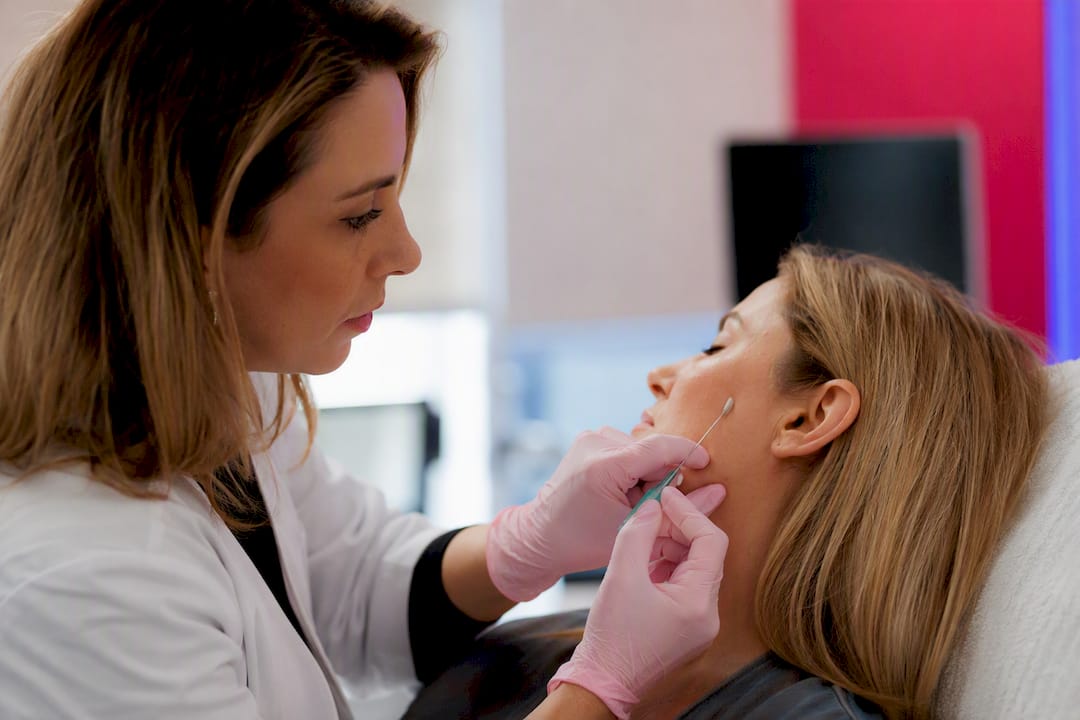
Needle injection, however, is not necessarily more painful when confined to a small, targeted area such as the vermilion border or a single glabellar rhytid. A short, precise needle pass over millimetres of tissue can feel comparable to, or even less noticeable than, a cannula’s longer subcutaneous passage, which some patients describe as a dragging or pressure sensation rather than a sharp point.
Topical or injectable anaesthesia narrows the gap further. Most modern fillers, including Belotero Balance formulations with lidocaine, reduce procedural pain regardless of delivery instrument, which means the comfort differential between cannula and needle often matters less than anaesthetic protocol and injection speed. Slower, more controlled delivery, whichever instrument is used, consistently correlates with lower reported pain scores in clinical practice observation, even without a large randomised trial to quantify it precisely.
The practical takeaway: recommend cannula technique to patients anxious about bruising and multiple puncture points across a broad zone, but don’t assume needle technique is automatically the more uncomfortable option for small, targeted corrections.
Technique protocols: how injection method differs by instrument
Needle technique typically follows serial puncture, linear threading, or fanning patterns, with the practitioner controlling depth and direction through direct, short passes. Aspiration before injection, while debated for its reliability with fine-bore needles, remains a common precaution, alongside slow, low-pressure delivery and constant needle repositioning to avoid depositing a large bolus in one location.
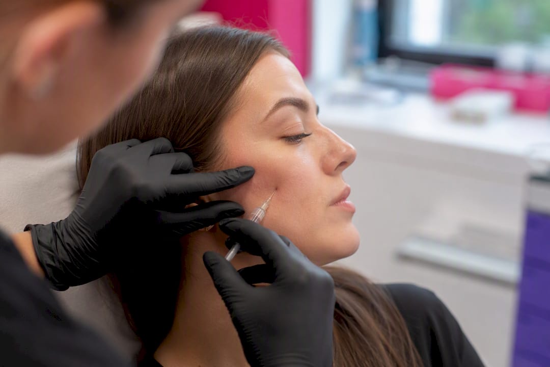
Cannula technique centres on a single entry point, usually created with a sharp needle “pilot hole,” through which the cannula threads to its target depth before fanning radially or delivering linear retrograde threads as it withdraws. Retrograde and anterograde threading each carry different backflow implications, and the tracer study on plane maintenance suggests retrograde technique with slower withdrawal reduces the backflow rate seen in faster passes.
Depth control differs meaningfully between the two. Needle depth is set at the point of insertion and rarely changes mid-pass; cannula depth can drift slightly as the flexible shaft travels through tissue planes of varying resistance, which is why experienced injectors periodically tent the skin to confirm the cannula tip’s position during longer passes.
Bolus size should generally run smaller with needle technique in vascular-dense zones, since a static needle position risks a larger volume being deposited at a single point if a vessel is inadvertently entered. Cannula technique tolerates slightly larger boluses per pass in appropriate compartments, given the reduced puncture risk, though the higher backflow rate found in tracer studies means practitioners should still favour multiple smaller passes over one large deposit.
Does instrument choice affect filler longevity or efficacy?
Filler longevity depends far more on product rheology, cross-linking technology, and injection depth than on whether a cannula or needle delivered it. A high G-prime filler placed correctly in the deep subcutaneous or supraperiosteal plane will last a comparable duration whichever instrument achieved that placement, provided the plane itself is accurate.
That last condition is where instrument choice does matter indirectly. Misplaced filler, whether too superficial or too deep, tends to migrate, show visibly, or resolve faster than intended regardless of which instrument placed it.
Needle technique’s precision advantage at a single point can actually support longevity in small, targeted zones, since accurate supraperiosteal placement against bone, difficult to achieve reliably with a flexible cannula shaft, gives structural fillers the stable foundation they need to persist. Products like EPTQ S300 used for fine contouring rely on that kind of placement accuracy more than on raw material volume.
There is no published head-to-head trial directly comparing filler longevity by instrument alone, holding product and technique constant, so any claim of one instrument producing categorically better results overstates the current evidence. The honest position is that correct placement, not instrument choice alone, drives how long a result lasts.
Which patient factors point towards one instrument over the other?
Certain patient presentations shift the calculation regardless of the practitioner’s usual preference. Patients on anticoagulant or antiplatelet therapy bruise and bleed more readily with either instrument, but the reduced puncture trauma of cannula technique generally produces less visible bruising in this group, making it the more considerate choice where anatomy allows.
Patients with a history of vascular events from previous filler treatments, anywhere on the face, warrant particular caution. A cannula in the previously affected zone reduces re-injury risk, though the underlying anatomical risk that caused the original event should be reassessed regardless of instrument.
Thin-skinned patients, common in the tear trough and perioral area, tend to show needle marks and bruising more visibly, another factor favouring cannula technique where the zone permits it. Conversely, patients seeking very precise, minimal-volume correction, such as a single deep nasolabial crease or a small perioral rhytid, are better served by a needle’s placement accuracy than by a cannula’s broader reach.
Scar tissue from previous surgery or trauma can restrict cannula passage, since the blunt tip struggles to navigate fibrous, non-uniform tissue planes. A needle’s rigidity often, in this specific circumstance, achieves more reliable placement, even in zones that would normally favour a cannula. Anatomical variation is common enough that pre-procedure palpation and, where relevant, ultrasound assessment should guide the final decision rather than a fixed zone-based rule alone.
Sterilisation and infection control: cannula vs needle considerations
Both instruments are single-use, sterile, disposable devices in modern aesthetic practice, and neither should ever be reused between patients or reinserted into a multi-dose vial after skin contact. That said, several practical differences affect infection control workflow.
Cannulas require a needle to create the initial skin entry point, meaning every cannula procedure involves two sterile devices rather than one. Both must be opened, checked for packaging integrity, and disposed of correctly as sharps waste, including the cannula itself despite its blunt tip, since the entry needle carries all the sharps risk.
Fewer skin punctures with cannula technique mean fewer breach points for surface bacteria to enter, a modest but real infection control advantage in procedures covering broad zones with multiple needle passes otherwise required. Strict skin antisepsis before the single entry point remains essential regardless, using an appropriate antiseptic solution and allowing it to dry fully before insertion.
Needle technique’s multiple puncture points, common with serial puncture or fanning patterns, each represent a discrete entry site requiring the same antiseptic standard. In practice, this means needle-heavy techniques demand more disciplined, repeated attention to antisepsis across a session rather than a single careful preparation.
Gloved, no-touch technique applies equally to both instruments. Neither cannula nor needle should contact any non-sterile surface, including the practitioner’s gloves at the fingertip, before insertion.
What the data actually tells practitioners to do
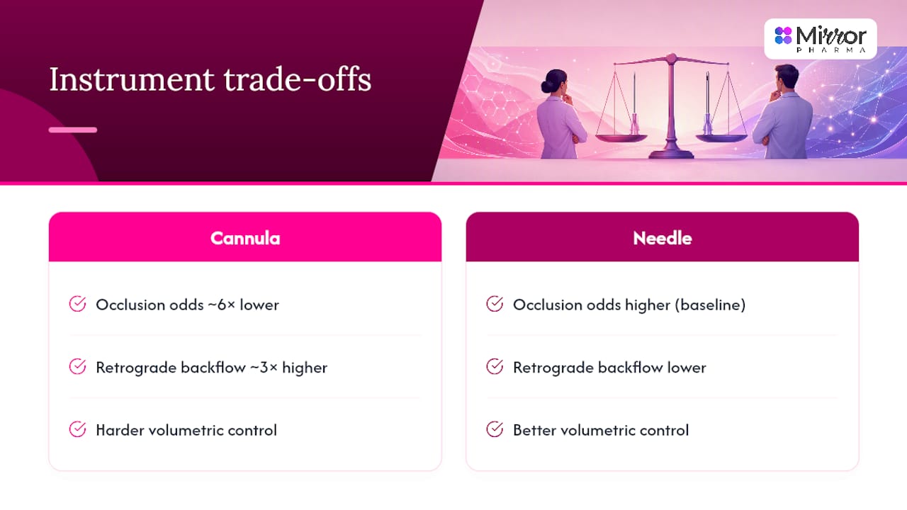
The conventional advice, “use a cannula for safety, a needle for precision,” is broadly correct but incomplete. What the cohort and tracer data actually support is more specific: cannulas lower occlusion odds by roughly sixfold in vascular-dense compartments, but they trade that safety margin for a threefold higher rate of retrograde backflow, meaning volumetric control gets harder exactly where the cannula is meant to help most.
Where the standard advice falls short is treating experience as a footnote rather than a primary variable. The same cohort data shows injector experience and case volume shift occlusion odds independently of instrument. A highly experienced needle injector working a familiar zone may carry lower risk than a novice cannula user working an unfamiliar one. Instrument choice matters, but it is not the only lever, and treating it as the sole safety decision oversimplifies what the evidence shows.
If you prioritise one thing from this guide, make it this: match instrument to anatomy first, then be honest about where your own supervised case volume sits before choosing the higher-risk option in a high-risk zone.
Sourcing cannulas, needles, and hyaluronidase for your clinic
Getting the right gauge of cannula or needle on the day it’s needed matters more than most procurement checklists admit, and running out of a specific bore mid-week forces compromises that shouldn’t be happening at the point of patient care. Mirror Pharma supplies qualified aesthetic professionals with cannulas, needles, dermal fillers, and hyaluronidase through verified, compliant ordering, with extended order cut-off times built specifically so clinics can match same-week stock to the actual cases on their schedule rather than whatever happens to be left in the stockroom.
For practitioners building volumising protocols around the zone-based recommendations covered above, the HyaCorp FACE and Belotero Volume with lidocaine ranges are available to order now, alongside the full consumables range needed to run a cannula-led or needle-led session safely. Check current stock and place your order through the Mirror Pharma product catalogue to have your next case fully equipped before it’s on the schedule.
This article is general information, not a substitute for advice from a qualified doctor. Consult a qualified healthcare professional about your own circumstances before acting on anything here.
Sources
- Precision in dermal filling: a comparison between needle and cannula using radiopaque tracers – PubMed
- Needles versus cannulas: advantages, disadvantages, and selection criteria for injecting dermal fillers – PubMed
FAQ
How long can a cannula stay on?
A cannula used for filler injection is a single-use device for one procedure only; it is discarded immediately after use and never reinserted or reused, including within the same session on a different patient.
Is cannula better for cheek fillers?
Yes, for most cheek volumisation cases, since the lateral and midface cheek involves a broad compartment and moderate vascular density where cannula technique’s plane maintenance and reduced puncture trauma offer a meaningful safety margin over needle-based fanning.
Is a cannula part of a needle?
No, they are distinct instruments; a cannula has a blunt, rounded tip and typically requires a separate sharp needle first to create the skin entry point it then passes through.
Is lip filler with a cannula safer?
Cannula technique can reduce bruising and vascular risk in lip volumisation, particularly for the body of the lip, but many practitioners still prefer a needle for precise definition along the vermilion border itself, making a hybrid approach common in practice.