

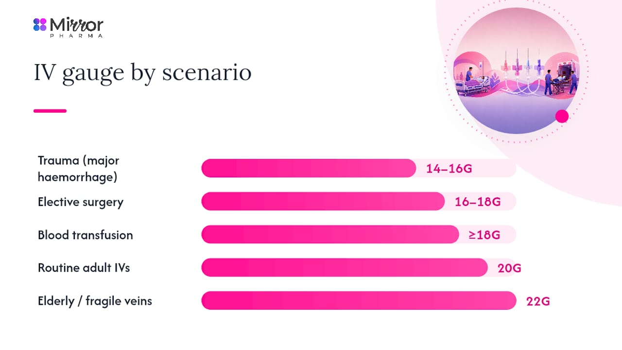
Choose the smallest gauge that will safely deliver the prescribed therapy. In practice, that means 14–16G for rapid resuscitation, 18G for blood transfusion and CT contrast, 20G for routine adult fluids and medications, and 22–24G for fragile, elderly, or paediatric veins. Peripheral IV cannulas range from 14G to 26G, with colour-coded hubs that follow ISO 6009 conventions — though colour alone is never sufficient to confirm gauge at the bedside.
Common IV cannula gauge → typical use at a glance:
| Gauge | Typical clinical use |
|---|---|
| 14G | Major trauma, rapid resuscitation, massive haemorrhage |
| 16G | High-volume infusion, surgical prep, emergency fluids |
| 18G | Blood transfusion, CT contrast, general surgery |
| 20G | Routine adult IV fluids and medications |
| 22G | Elderly, fragile veins, low-flow medications |
| 24G | Paediatrics, neonates, very fragile adult veins |
| 26G | Neonates, specialist small-vessel access |
- 14–16G are the go-to choices when speed of delivery is the priority.
- 18G is the minimum recommended for blood transfusion and contrast injection.
- 20G covers the majority of adult ward infusions without unnecessary vein trauma.
- 22–24G suits paediatric and neonatal patients, and adults with difficult or fragile access.
Procurement note: always verify the gauge printed on the manufacturer’s pack label. Colour coding is a guide, not a guarantee — stock from different suppliers can vary.
Key takeaways
| Point | Details |
|---|---|
| Smallest effective gauge | Choose the narrowest cannula that meets the therapy’s flow and viscosity requirements to reduce vein trauma. |
| Scenario mapping | 14–16G for resuscitation; 18G for transfusion and contrast; 20G for routine adult fluids; 22–24G for paediatrics and fragile veins. |
| Catheter-to-vessel ratio | Keep the catheter’s outer diameter at or below approximately 45% of the vein lumen to maximise dwell time and reduce phlebitis. |
| Length matters | A catheter too short for a deep vein predisposes to early dislodgement; match length to vein depth and site. |
| Mirror Pharma | Supplies IV dressings, infusion therapy products, and clinical consumables to verified healthcare professionals via its online shop. |
How are IV cannula sizes measured?
The standard measurement system for peripheral IV cannulas is the Birmingham Wire Gauge (BWG), commonly called simply “gauge” or “G.” The counterintuitive rule is that a higher gauge number means a narrower lumen. A 14G cannula has a substantially larger outer diameter than a 22G. NCBI Bookshelf confirms this inverse relationship and notes that manufacturers vary in catheter length and exact outer diameters, so the figures below are approximate.

The French system uses a different scale: French size = outer circumference in millimetres, so French size divided by π gives the outer diameter. It is primarily used for larger central venous catheters, chest drains, and urinary catheters. For routine peripheral IV cannulas, the French system is not applied — gauge is the universal reference. If you encounter French sizing on a peripheral device, check the device instructions for use (IFU) carefully, as it is outside normal convention.
Manufacturer variance matters more than many students expect. Two 20G cannulas from different suppliers can differ by 0.1–0.2 mm in outer diameter, which affects catheter-to-vessel ratio calculations in smaller veins. When stocking or ordering, check the device IFU for the exact specification rather than relying on gauge alone.
What do cannula hub colours actually mean?
ISO 6009 establishes the colour coding most clinicians recognise on cannula hubs and packaging. The table below shows the standard mapping alongside approximate outer diameters and typical uses.
Pro Tip: Never select a cannula based on hub colour alone. Hospital stock from different manufacturers occasionally deviates from ISO 6009, and a colour mismatch between suppliers has caused clinical errors. Always read the gauge printed on the packet before opening.
Less common sizes worth knowing: 26G devices are stocked in neonatal and specialist paediatric units but rarely appear on general wards. Long peripheral catheters (sometimes called “long lines” or “extended dwell” catheters) are available in 18G and 20G and are used when a standard 25–32 mm catheter cannot reach a stable vein segment, particularly in patients with obesity or difficult access. These are distinct from midlines and PICCs and carry their own insertion and dwell-time considerations.
What flow rates can you expect from each gauge?
Flow rate is where gauge choice becomes clinically consequential. The figures below are approximate gravity-fed rates; pressure bags and infusion pumps alter these substantially. Published flow rate reference data provides the following typical values for water at room temperature.
These figures assume water viscosity and a standard catheter length. Blood is roughly three to four times more viscous than water, so transfusion flow through an 18G will be considerably lower than the table suggests for saline. Similarly, contrast media used in CT is viscous and often warm-injected under power; most CT protocols specify a minimum of 18G and prefer 16G for high-flow injections.
Clinical implication: A 20G cannula delivers roughly 65 mL/min under gravity — adequate for most maintenance infusions but insufficient for rapid volume resuscitation. When a patient needs fluid fast, the gauge is not a minor detail.
Flow rate also depends on catheter length: a longer catheter of the same gauge produces more resistance and lower flow. Real-world flow depends on device length, fluid viscosity, and whether a pressure device is in use, so local testing and device IFUs remain the definitive reference for your specific stock.
Which cannula size should you use for common clinical scenarios?
MSD Manuals recommends 18–20G for routine adult infusions, 14–16G for high-volume infusion, and 22–24G for infants and small children. The scenario-led breakdown below adds clinical rationale to those recommendations.
- Trauma and major haemorrhage: 14–16G, two large-bore peripheral lines. Speed of delivery is the priority; follow your institution’s massive transfusion protocol.
- Elective surgery and surgical prep: 16–18G. Allows rapid fluid boluses intraoperatively without the vein trauma of a 14G in a non-emergency.
- Blood transfusion: 18G minimum. Smaller gauges increase haemolysis risk and slow delivery unacceptably for most transfusion schedules.
- CT contrast injection: 18G minimum; 16G preferred for high-flow power injection. Check the scanner protocol — many specify a minimum flow rate that a 20G cannot reliably meet.
- Routine adult IV fluids and medications: 20G. Covers the vast majority of ward infusions with minimal vein trauma.
- Elderly patients or fragile veins: 22G. Prioritise vein preservation; a 20G in a fragile vein causes more phlebitis and earlier failure.
- Paediatrics (school age and older): 22–24G depending on vein size and therapy.
- Neonates and infants: 24–26G. Specialist paediatric and neonatal units will have local protocols.
Decision checklist before choosing gauge:
- What is the therapy, and what flow rate does it require?
- Is the fluid viscous (blood, contrast, lipid emulsion)?
- How urgent is delivery?
- What is the vein quality — size, depth, fragility?
- What is the expected dwell time?
- Does a local protocol (transfusion, contrast, massive haemorrhage) specify a minimum gauge?
When a protocol exists, follow it. Institutional policies on transfusion and contrast are written around evidence and equipment-specific flow data that a general guide cannot replicate.
How to decide cannula size: a bedside checklist
A repeatable decision sequence prevents the common error of defaulting to whatever is in the trolley.
- Read the prescription or order. Identify the fluid or drug, the required rate, and any protocol-specified gauge.
- Estimate required flow. Cross-reference with the flow rate table above; factor in viscosity if relevant.
- Inspect and palpate the vein. Assess size, depth, and fragility before committing to a gauge.
- Apply the catheter-to-vessel ratio rule. The catheter’s outer diameter should occupy no more than approximately 45% of the vein’s estimated lumen. A catheter that fills more than half the vein restricts blood flow around the device, accelerating phlebitis and reducing dwell time.
- Select gauge and length. Smallest gauge that meets the therapy; longest length that sits comfortably within the chosen vein segment.
- Plan for escalation. If the vein is inadequate for the required gauge, document and escalate — do not force a large-bore cannula into a small vein.
Pro Tip: A useful bedside rule: if you need rapid infusion or blood, use 18G or larger; if the vein is small, default to 20G; treat a running 24G as acceptable only when therapy is genuinely low-flow. Write the gauge and rationale in the notes.
Document the chosen gauge, insertion site, date, time, and clinical indication. This supports escalation decisions and is required by most institutional policies.
Does catheter length affect how a cannula performs?
Yes, and it is underappreciated by many students. A catheter that is too short for the chosen vein sits with its tip near the insertion point rather than in a stable vein segment, predisposing to early dislodgement and infiltration. One that is too long for a small vein risks perforation of the posterior wall.
Common catheter lengths by gauge and context:
- 14–16G: typically 45–50 mm. Designed for large antecubital or forearm veins where a longer track is needed to maintain stability under high-flow conditions.
- 18–20G: typically 32–45 mm. Standard adult peripheral length; suits most forearm and antecubital sites.
- 22G: typically 25–32 mm. Shorter to match the smaller veins it is placed in.
- 24–26G: typically 19–25 mm. Neonatal and paediatric lengths; some adult 24G devices are available at 19 mm for very superficial veins.
When a standard-length catheter cannot reach a stable vein segment, the options are a long peripheral catheter (typically 6–15 cm, inserted under ultrasound guidance into a deeper forearm vein), a midline catheter (8–20 cm, tip in the proximal basilic or cephalic vein), or escalation to a PICC for longer-term access. The decision to escalate should be made before multiple failed attempts damage the vein network. VeinCraft Academy’s catheter size selection guidance notes that longer catheters are often needed for deeper veins or patients with obesity to ensure the tip sits in a healthy vein segment, and that short catheters in deep veins predispose to early dislodgement.
Key technique points that affect cannula function and longevity
Gauge selection is only part of the picture. How the cannula goes in determines whether it stays in.
- Skin preparation. Clean the site with a 2-ply alcohol swab and allow it to dry fully before insertion. Wet skin reduces antiseptic efficacy and increases infection risk.
- Vein stabilisation. Apply distal traction to anchor the vein. A mobile vein rolls away from the needle and is the most common cause of failed first attempts.
- Entry angle. 10–30 degrees for superficial veins; a steeper angle risks posterior wall puncture. Reduce the angle once the needle is in the vein.
- Bevel orientation. Bevel up for initial entry; this reduces the force needed to penetrate the skin and vein wall.
- Flashback confirmation. Wait for blood in the flashback chamber before advancing the catheter. Advancing without flashback is the primary mechanism of infiltration.
- Catheter advancement. Advance the plastic catheter off the needle with a smooth, continuous motion. Partial advancement leaves the tip near the vein wall and increases the risk of positional occlusion.
- Securement. Apply a sterile transparent film dressing (such as 3M Tegaderm) over the insertion site. For sites crossing a joint, consider a splint to reduce movement-related dislodgement. Check for leakage at the hub after flushing.
Pro Tip: Document the number of insertion attempts, the site, the gauge, and the date. If a second clinician needs to remove or replace the cannula, this information directly informs their approach and supports escalation decisions.
For patients with needle anxiety or thin skin, topical anaesthetic cream applied 45–60 minutes before insertion can improve cooperation and reduce vein spasm, particularly in paediatric patients.
Common complications linked to cannula size and how to reduce them
Size-related complications are largely preventable when gauge and length are chosen deliberately.
- Phlebitis (vein inflammation): the most common complication. A catheter that is too large for the vein restricts blood flow around the device, increasing endothelial shear and inflammatory response. Mitigate by choosing the smallest effective gauge, rotating sites every 72–96 hours per local policy, and inspecting the site at every drug round.
- Infiltration (fluid into surrounding tissue): occurs when the catheter tip exits the vein lumen. More common with short catheters in mobile veins. Check for swelling, coolness, and slowed infusion rate. Stop the infusion immediately if infiltration is suspected.
- Extravasation (vesicant fluid into tissue): a subset of infiltration involving drugs that cause tissue necrosis. Requires immediate cessation, aspiration if possible, and escalation to the vascular access team or pharmacy for antidote guidance. Never continue an infusion through a cannula with any doubt about patency.
- Haemolysis during transfusion: using a gauge smaller than 18G for blood transfusion can cause red cell destruction through mechanical shear. Use 18G or larger for all routine transfusions.
- Thrombosis and thrombophlebitis: larger catheters in smaller veins, prolonged dwell, and irritant drugs all increase risk. Minimise by matching catheter size to vein size and reviewing the cannula daily for continued clinical necessity.
Red flags requiring immediate action: pain or burning during infusion; visible swelling or induration at the site; resistance when flushing; skin blanching or discolouration around the insertion point; patient reporting a change in sensation. Any of these signs warrants stopping the infusion, assessing the site, and removing the cannula if in doubt.
Document any complication, the action taken, and the outcome. Extravasation of a vesicant drug and any suspected catheter-related bloodstream infection should be reported through your institution’s incident reporting system and escalated to a senior clinician or vascular access team.
Evidence snapshot: flow rate, shear stress and catheter-to-vessel ratio
The bedside rules in this guide are not arbitrary. They are grounded in a small but consistent body of evidence.
- Catheter-to-vessel ratio: VeinCraft Academy’s size selection guidance, drawing on Infusion Nurses Society standards, recommends keeping the catheter’s outer diameter at or below approximately 45% of the vein’s estimated lumen diameter. Above this threshold, blood flow around the catheter is restricted, endothelial shear increases, and both phlebitis rates and early failure rates rise.
- Flow rate evidence: the Wolters Kluwer flow rate reference guide provides the gauge-to-flow figures used in this article and notes that real-world flow depends on device length, fluid viscosity, and pressure devices — published figures are anchors, not guarantees.
- Gauge and diameter physiology: NCBI Bookshelf confirms the inverse gauge-to-diameter relationship and explains why the French system is not applied to routine peripheral lines.
- Clinical gauge selection: MSD Manuals provides the clinical gauge ranges (14–16G for high-volume, 18–20G for routine, 22–24G for paediatrics) that underpin the scenario recommendations above.
Bedside translation: a catheter occupying more than half the vein lumen is too large for that vein, regardless of whether the gauge is technically appropriate for the therapy. If the vein cannot accommodate the required gauge without exceeding the 45% ratio, escalate to a larger vein or a different access device.
The practical implication is straightforward: choose the smallest gauge that delivers the therapy, placed in the largest accessible vein appropriate for that gauge. This combination maximises dwell time, minimises phlebitis, and reduces the need for re-siting.
Practical bedside tips and common pitfalls
Do:
- Confirm the gauge on the packet label before opening, every time.
- Use ultrasound for difficult or deep venous access rather than making multiple blind attempts.
- Flush with 0.9% sodium chloride before and after each drug administration to confirm patency and reduce drug interaction at the hub.
- Reassess the cannula daily for clinical necessity, signs of phlebitis, and site integrity.
- Consider topical anaesthetic (such as EMLA cream) for needle-anxious patients or children.
Don’t:
- Upsize the gauge unnecessarily. A 14G in a routine maintenance patient causes more vein trauma and more pain than a 20G, with no clinical benefit.
- Rely on hub colour alone to confirm gauge.
- Advance the catheter without clear flashback.
- Leave a cannula in situ beyond its clinical indication or beyond your institution’s maximum dwell-time policy.
Three quick checks before flushing or starting an infusion:
- Aspirate gently for blood return to confirm intravascular placement.
- Flush with 2–5 mL of 0.9% sodium chloride and observe the site for swelling or resistance.
- Confirm the infusion rate and drug compatibility before connecting the giving set.
Pro Tip: If you encounter resistance during flushing, stop immediately. Do not apply force. A blocked or misplaced cannula that is flushed forcefully can cause extravasation or catheter embolism. Remove and re-site.
A note on intended audience and supervised practice
This guide is written for licensed practitioners, qualified healthcare professionals, and students in supervised clinical training. The gauge recommendations, flow rate figures, and decision checklists here are educational references, not a substitute for institutional protocols or direct clinical supervision.
Cannula insertion is a procedural skill that requires hands-on training and sign-off under a qualified supervisor. If you are a student or trainee, use this material to prepare for supervised practice and to understand the rationale behind the choices your supervisors make. Always defer to local guidelines, your institution’s vascular access policy, and the device IFU for the specific product in use. Training academies and clinical educators are encouraged to use this material as a structured reference alongside practical skills sessions.
Mirror Pharma: consumables and infusion products for qualified practitioners
For licensed practitioners and aesthetic clinics sourcing IV consumables and infusion therapy products, Mirror Pharma offers a straightforward B2B supply route with extended order cut-off times that suit busy clinical schedules. The shop stocks a range of products relevant to infusion practice, including IV dressings, vitamins, and infusion therapy products alongside the broader aesthetic and pharmaceutical range. All products are supplied to verified healthcare professionals and prescribers. Browse the Mirror Pharma shop to check current stock and place an order.
Sources
- NCBI Bookshelf
- MSD Manuals — How to insert a peripheral intravenous catheter
- IV catheter size and flow rate reference guide (Wolters Kluwer content hub)
- VeinCraft Academy — IV catheter size selection guide
Consult your institution’s vascular access policy and the device IFU for the specific cannula you are using. Local guidelines take precedence over any general reference for procurement, stocking, and clinical decision-making.
FAQ
What are the standard sizes of IV cannulas?
Peripheral IV cannulas commonly range from larger to smaller gauges in clinical practice. The most commonly used sizes in adult clinical practice are 18G, 20G, and 22G, with 14–16G reserved for high-volume resuscitation and 24–26G for neonatal and paediatric use.
Is a 14G or 20G cannula bigger?
A 14G cannula is larger. In the gauge system, a lower number means a wider outer diameter — 14G has an outer diameter of approximately 2.0–2.2 mm, compared with approximately 1.0–1.1 mm for a 20G.
How do you decide which cannula size to use?
Follow any institutional protocol that specifies a minimum gauge, such as transfusion or CT contrast policies.
What are the common IV cannula sizes used in UK clinical practice?
The most frequently stocked sizes in UK hospitals are 18G (green), 20G (pink), and 22G (blue), following ISO 6009 colour coding. Larger 14G and 16G devices are held in emergency and theatre settings; 24G is standard in paediatric and neonatal units.