
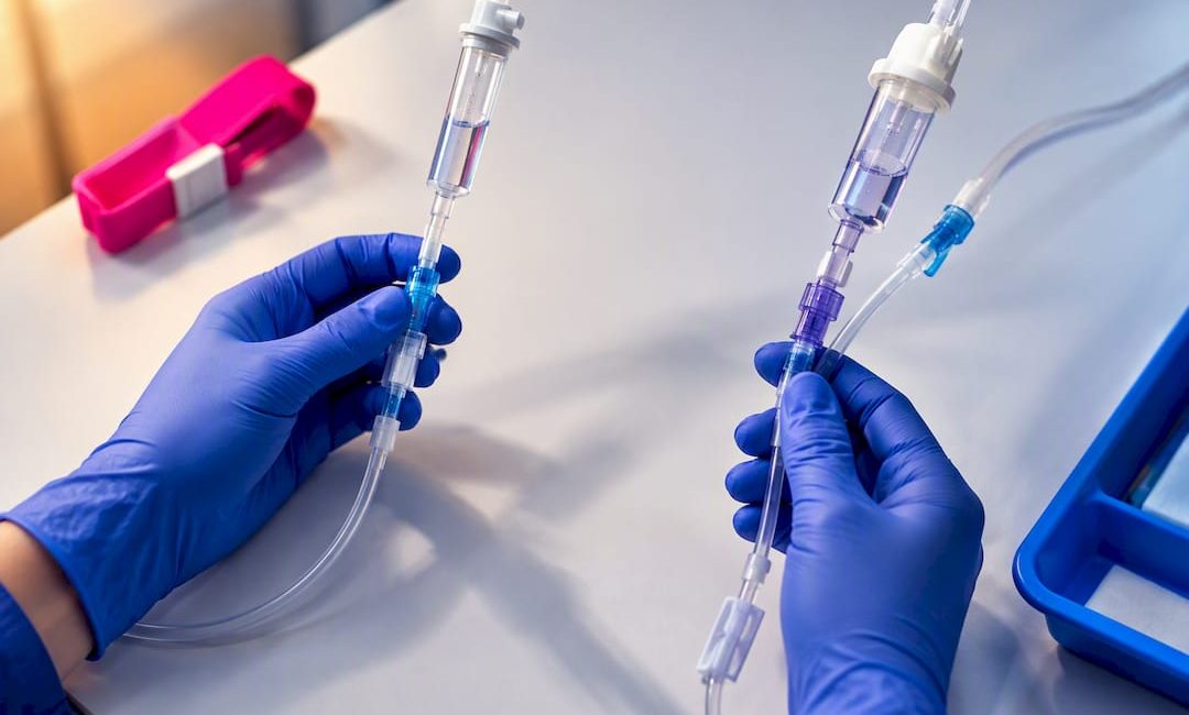
Infusion lines split into peripheral devices (short peripheral cannulae and midlines) and central devices (PICC, nontunnelled and tunnelled central venous catheters, and implanted ports). Peripheral access suits short, low-risk infusions, while central access is reserved for long-term therapy, vesicant drugs or parenteral nutrition. Administration sets add a second layer of choice: primary and secondary tubing, macro-drip and micro-drip, vented and non-vented.
TL;DR:
- Central devices like PICCs, tunnelled lines, and ports are suitable for long-term therapy, vesicant drugs, or parenteral nutrition, with tip placement confirmed in the lower superior vena cava.
- Midline catheters should not be used for vesicants or high-osmolar solutions, and timely transition to central access is crucial if therapy duration exceeds their practical limit.
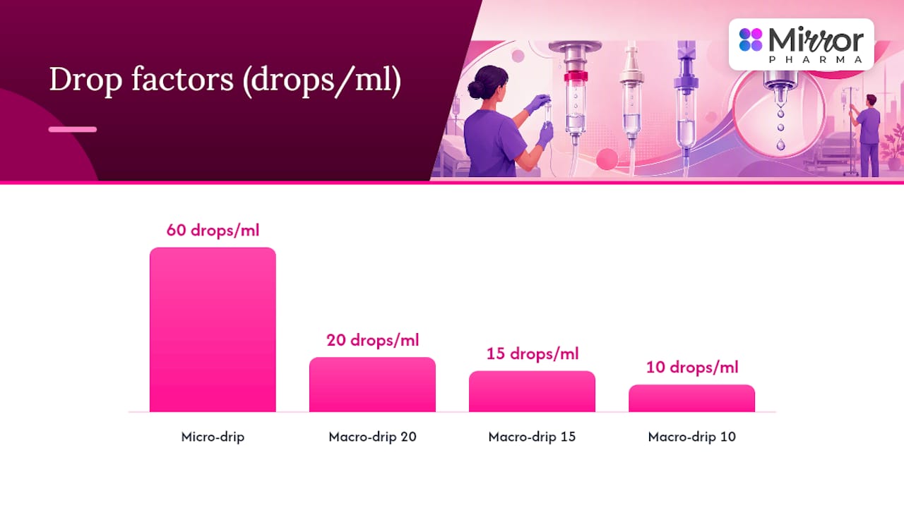
- Drop factors must be verified before infusion rate calculation, with micro-drip sets delivering 60 drops per milliliter and macro-drip sets delivering 10, 15, or 20 drops.
- Ultrasound guidance is now standard for central line insertion, and strict adherence to aseptic techniques reduces complications like infection and mechanical failures.
- Regular device assessment, proper flushing with push-pause technique, and prompt removal when no longer necessary minimize complications and improve patient safety.
Table of Contents
- Overview: taxonomy of vascular access devices and administration sets
- Peripheral devices: short peripheral IV catheters and midline catheters
- Central devices: PICC, nontunnelled CVC, tunnelled CVC and implanted ports
- Infusion sets and tubing: primary, secondary and specialised options
- Selecting the right line: a bedside decision framework
- Insertion and maintenance best practice: reducing complications
- Complications and troubleshooting: recognising trouble early
- Practice implications for licensed practitioners
- How Mirror Pharma supports safe infusion practice
- Sources
- FAQ
Overview: taxonomy of vascular access devices and administration sets
Vascular access devices fall into two broad families, and the distinction shapes almost every decision that follows. Peripheral devices sit in a vein close to the skin surface, typically in the arm or hand, and are suited to therapies that are short in duration and gentle on the vessel wall. Central devices thread a catheter into one of the great veins so that the tip finishes near the heart, which matters because that location dilutes irritant drugs quickly and tolerates therapy that would damage a smaller peripheral vein.
Tip position is not a technicality. According to vascular access guidelines from NHS Highland, a central catheter tip should sit in the lower third of the superior vena cava or the upper right atrium, while midline catheters, despite their length, still terminate in a peripheral vein and are classed accordingly. Getting this wrong on a chest X-ray reading, or failing to confirm it after insertion, is one of the more consequential documentation gaps in vascular access practice.
Administration sets form the second half of the taxonomy. Primary sets connect directly to the vascular access device and deliver the main infusion; secondary sets, often called piggyback lines, connect into an injection port on the primary line to deliver an intermittent medication before the primary fluid resumes. Within these categories, sets are further defined by drop factor and by whether they are vented or non-vented.
The chapter on IV therapy management from the NCBI Bookshelf confirms this primary and secondary structure and adds a third dimension: drip chamber design. Macro-drip chambers deliver larger, countable drops and are the default for most adult infusions, while micro-drip chambers deliver much finer drops and are reserved chiefly for paediatric or precise low-volume delivery.
Arterial cannulae deserve a brief mention here because they are easily confused with venous devices on a busy ward round. An arterial line accesses a peripheral artery, usually the radial, for continuous blood pressure monitoring or blood gas sampling rather than for drug delivery. It is never used to infuse medication, and mistaking one for a venous line is a recognised, serious safety event rather than a semantic slip.
Peripheral devices: short peripheral IV catheters and midline catheters
Short peripheral IV catheters, commonly called PIVs or cannulae, are the workhorse of everyday infusion therapy. They are inserted into superficial veins on the dorsum of the hand, the forearm or occasionally the antecubital fossa, using gauges chosen to match both the vein and the therapy: finer gauges for fragile or small veins, wider gauges when rapid flow rates or viscous fluids are anticipated. Dwell time is short by design, and ongoing guidance now favours removing a PIV when it is clinically indicated rather than swapping it on a fixed calendar schedule, a shift confirmed in WHO guidance on catheter-related infection prevention.
Midline catheters occupy the space between a standard cannula and a central line. They are longer, typically measuring 7.5 to 20 centimetres according to the NHS Highland vascular access guidelines, and the tip still ends in a peripheral vein rather than near the heart. That makes a midline a peripheral device by classification even though its insertion technique and dwell expectations look closer to central practice. Midlines are chosen when treatment is expected to run for up to a few weeks, when preserving the patient’s remaining peripheral veins matters, or when repeated cannulation attempts have made standard PIV access difficult.
Midlines are not a substitute for central access when the drug itself demands it. They must not be used for continuous vesicant infusions, for high-osmolarity parenteral nutrition or for solutions at the extremes of pH, a restriction the quality improvement literature review on peripheral catheters applies to peripheral devices generally and which extends to midlines by the same vessel-protection logic.
Practical care points for both device types:
- Flush with a minimum 10 ml syringe using a push-pause technique to clear the lumen without generating excessive pressure.
- Prime needle-free access devices with 0.9% sodium chloride before first use and check patency before every infusion.
- Secure the device well and inspect the insertion site at every shift for redness, swelling or leakage.
- Escalate to central access promptly if the expected duration extends beyond a midline’s practical window or if a vesicant drug becomes necessary.
Pro Tip: If a midline dwell is approaching its planned end and therapy is set to continue, plan the switch to central access before the vein becomes compromised, not after.
Central devices: PICC, nontunnelled CVC, tunnelled CVC and implanted ports
Peripherally inserted central catheters, or PICCs, are inserted through a vein in the upper arm, usually the basilic, brachial or cephalic vein, and threaded until the tip reaches the lower superior vena cava. They are available in single, double or triple lumen configurations depending on how many simultaneous infusions the patient needs, and most PICCs carry external markings that let staff confirm whether the catheter has migrated inward or outward since insertion. Dwell times commonly extend from several weeks to a few months, which makes a PICC the practical choice for extended courses of intravenous antibiotics or chemotherapy that fall short of needing a permanent implanted device.
Nontunnelled central venous catheters are inserted directly into a large vein, typically the internal jugular, subclavian or femoral, with no subcutaneous tunnel between the skin entry point and the vein. They are the fastest route to central access and are common in critical care and emergency settings, but that speed carries a trade-off: a higher relative risk of mechanical complication at insertion and a dressing regimen that needs close attention because the exit site sits directly over the vein.
Tunnelled catheters and implanted ports both route the catheter under the skin before it enters the vein, which anchors the device and moves the exit site away from direct vessel access.
- Tunnelled central venous catheters exit through the skin at a distance from the venipuncture site and suit patients needing weeks to months of frequent access, such as those on long courses of chemotherapy or home parenteral nutrition.
- Implanted ports sit entirely under the skin with no external component between uses, accessed through a needle puncture into a subcutaneous reservoir, and are the preferred choice when access will be needed intermittently over months or years.
- Both device types demand imaging confirmation of tip position before first use, following the same lower superior vena cava or right atrial target used for other central lines.
- Care differs sharply from PICCs and nontunnelled lines: ports need no daily dressing between accesses, while tunnelled lines still require routine exit-site care.
Ultrasound guidance has become the expected standard for central line insertion rather than an optional refinement. Paediatric CVAD guidance notes that ultrasound-guided insertion reduces mechanical complications compared with landmark-based technique, and the same logic applies across adult practice: visualising the vein and needle in real time cuts down on failed passes, arterial puncture and pneumothorax risk.
Infusion sets and tubing: primary, secondary and specialised options
Choosing the right administration set is as consequential as choosing the right catheter, and it is where small errors most often creep in. A primary set runs continuously from the fluid container to the vascular access device, while a secondary set piggybacks into a port on the primary line to deliver an intermittent drug, after which the primary fluid resumes automatically once the secondary bag empties.
Drop factor is the detail most likely to be assumed rather than checked. The chapter on IV therapy management sets out the standard split: macro-drip sets deliver 10, 15 or 20 drops per millilitre and are used for most adult gravity infusions, while micro-drip sets deliver 60 drops per millilitre and are reserved largely for paediatric infusions or situations demanding very fine control over a low flow rate.
Statistic callout: micro-drip sets deliver 60 drops per millilitre against 10 to 20 for standard macro-drip tubing. That sixfold difference in drop size means a rate calculation done with the wrong set in mind can be wrong by a similar margin, which is why the drop factor printed on the tubing packaging should be confirmed before any manual rate calculation, not assumed from habit.
Vented and non-vented sets are matched to container type rather than personal preference. A vented set allows air to enter a rigid glass or hard plastic container as fluid leaves it, preventing a vacuum that would otherwise stop the flow, while a non-vented set is designed for flexible plastic bags that collapse as they empty and need no air inlet. Specialised sets extend the same logic further: blood administration sets carry an in-line filter to trap debris and clots, and lightproof tubing protects light-sensitive drugs from degradation during delivery.
Practical steps that reduce common set-up errors:
- Confirm the drop factor printed on the packaging before calculating any gravity infusion rate.
- Fill the drip chamber to between a quarter and a half full, since an overfilled or underbilled chamber makes the drop count unreliable.
- Match vented tubing to rigid containers and non-vented tubing to collapsible bags before spiking the fluid.
- Recheck the clamp position and flow rate after any patient repositioning or line manipulation.
The same NCBI Bookshelf chapter flags failure to confirm the drop factor as a recurring cause of rate miscalculation in gravity infusions, a reminder that pump delivery removes some manual arithmetic but never removes the need to select the correct set and container pairing in the first place.
Selecting the right line: a bedside decision framework
The choice between peripheral and central access, and between the device types within each family, comes down to a small number of clinical axes rather than a long checklist.
- Duration of therapy: a course expected to last days favours a standard PIV, weeks favours a midline or PICC, and months to years favours a tunnelled line or port.
- Drug properties: vesicants, high-osmolarity parenteral nutrition and solutions at extreme pH values need central access because peripheral and midline veins cannot tolerate them safely, a limit set out in the peripheral catheter literature review.
- Venous access quality: a patient with poor or repeatedly exhausted peripheral veins is a candidate for a midline or PICC even when the drug itself would tolerate peripheral delivery.
- Number of concurrent infusions: patients needing several simultaneous incompatible drugs benefit from a multi-lumen PICC or CVC rather than juggling one lumen.
Rules of thumb follow from these axes reasonably directly. A standard PIV is right for routine short-term fluids and antibiotics measured in days. A midline earns its place when therapy will run for up to a few weeks and the drug is peripherally compatible. A PICC becomes the better fit once therapy stretches beyond a few weeks or multiple lumens are needed. A tunnelled line or port is reserved for therapy measured in months or years, particularly chemotherapy, home parenteral nutrition or other recurring long-term treatment.
Red flags that should prompt an immediate move to central access include any request for a vesicant infusion through a peripheral line, visibly poor or thrombosed peripheral veins, and a therapy plan that already anticipates weeks of parenteral nutrition. Whichever device is chosen, document the indication, the vein and device used, the confirmed tip position where relevant, and the planned review date, since that record is what lets the next clinician on shift make a safe decision without repeating the assessment from scratch.
Insertion and maintenance best practice: reducing complications
Good outcomes in vascular access depend less on the device chosen and more on how consistently it is inserted and maintained. Ultrasound guidance for central line insertion is now the evidence-based standard rather than an occasional aid, reducing mechanical complications compared with landmark technique according to paediatric CVAD guidance, and the same principle of visualising the target before committing a needle applies to difficult peripheral access too.
Aseptic non-touch technique, ANTT, governs every point of contact with the device: insertion, dressing changes and needle-free access. The vascular access devices clinical guidance sets out device-specific protocols that include chlorhexidine-impregnated dressings for central devices changed every 7 days, or sooner if the dressing becomes loose, damp or visibly soiled, and priming of needle-free access devices with 0.9% sodium chloride before use.
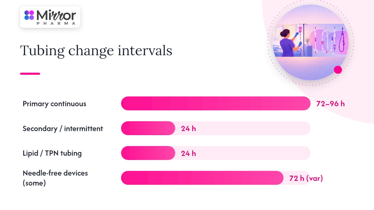
Tubing change intervals follow their own schedule, separate from dressing changes:
- Primary continuous tubing is commonly changed every 72 to 96 hours according to clinical procedures guidance on IV fluids and tubing.
- Secondary or intermittent tubing is changed more frequently, typically every 24 hours.
- Tubing carrying fat emulsions or parenteral nutrition is changed every 24 hours regardless of whether it is primary or secondary, because lipid residue supports microbial growth faster than clear fluids do.
- Needle-free access devices are changed per manufacturer instructions or local protocol, with some units specifying a 72-hour change interval in line with vascular access devices clinical guidance.
Flushing technique matters as much as flushing frequency. A minimum 10 ml Leur lock syringe should be used to flush or aspirate midline and central lumens, since smaller syringes generate disproportionately high pressure inside the lumen, a point made explicitly in the midline peripheral venous catheter protocol. A push-pause technique, alternating short bursts of flush with brief pauses, clears debris from the lumen wall more effectively than a single steady push.
Peripheral catheter guidance has moved firmly away from routine, calendar-based replacement. The WHO guidance on catheter-related infection prevention recommends daily site assessment and removal when clinically indicated, rather than swapping a functioning, uninfected line simply because a fixed number of hours has passed.
Pro Tip: Document every flush, dressing change and site assessment at the time it happens rather than at the end of the shift; a gap in the record is often the first thing an infection review will flag.
Know when a device has done its job. Remove any line as soon as it is no longer clinically necessary, since dwell time itself is a risk factor independent of how well the device has been cared for.
Complications and troubleshooting: recognising trouble early
Most serious vascular access complications give some warning if the line is being assessed properly, and the response follows a broadly consistent pattern.
- Occlusion: resistance on flushing or a sluggish infusion suggests a blocked lumen. Try a gentle push-pause flush with saline first; if that fails, enzymatic therapy such as urokinase may be required under prescription and local protocol, as described in the midline peripheral venous catheter protocol.
- Suspected catheter-related bloodstream infection: fever, chills or a visibly inflamed exit site without another obvious source should prompt cultures, review of dressing integrity and discussion of whether the device needs removing.
- Extravasation: swelling, pain or blanching around a peripheral or midline site during infusion means stopping the infusion immediately, disconnecting the line without flushing further fluid into the tissue, and following local protocol for the specific drug involved.
- Arterial cannulation: pulsatile, bright red backflow on insertion signals an artery rather than a vein. Withdraw the device, apply firm direct pressure for several minutes, and escalate for review before any further attempt.
- Air embolism: sudden breathlessness, chest pain or a drop in oxygen saturation during line connection or disconnection is an emergency. Clamp the line, position the patient appropriately and call for immediate medical assistance.
Prevention threads through all of these: an intact, dry dressing, correct needle-free device handling, confirmed tip position for central lines, and prompt removal of any device that is no longer needed all reduce the odds of reaching this list in the first place.
Practice implications for licensed practitioners
Vascular access decisions sit close to patient safety, which is why this guide is written for licensed practitioners, registered nurses and prescribers rather than for general readers. The devices, flush volumes and change intervals described here reflect published guideline sources, but individual patient factors, local infection control policy and specialist vascular access advice should always take precedence over a general article.
Mirror Pharma’s own IV vitamin therapy safety toolkit is built around the same maintenance principles covered above, aimed at practitioners running IV vitamin infusions in clinic. Anyone applying the decision framework in this guide to an individual patient should still confirm the plan against local policy and, where the picture is unclear, seek advice from a vascular access specialist rather than relying on generalised guidance alone.
— Rizwan
How Mirror Pharma supports safe infusion practice
Reliable infusion practice depends on having the right consumables on hand when a clinic list is running, not on hunting for stock between patients. Mirror Pharma supplies needles and consumables alongside IM/IV vitamin products to verified healthcare professionals, with extended order cut-off times built specifically so that practitioners running same-week clinics can order later in the day and still receive stock in time.
Access to the site is verified for licensed practitioners and qualified aesthetic professionals, in keeping with the prescription-only nature of many of the products involved. The IV vitamin therapy safety toolkit gives clinicians a practical reference for the same maintenance and flushing standards covered in this guide, built specifically for infusion practice in clinic rather than hospital settings.
None of this replaces individual clinical judgement or local infection control policy, and nothing here should be read as promotion of a prescription medicine to the general public. For practitioners who are verified and ready to order, the needles and consumables range and IM/IV vitamins category are the places to start.
Sources
- Vascular access in adults guidelines (Right Decisions / NHS Highland)
- Chapter 23 IV therapy management (NCBI Bookshelf)
FAQ
What are the three types of infusions?
Infusions are generally grouped as continuous, intermittent and bolus, referring to how the fluid or drug is delivered over time rather than to the device used. A continuous infusion runs steadily, an intermittent infusion runs on a schedule with breaks between doses, and a bolus delivers a set volume quickly. The device chosen, whether peripheral or central, is a separate decision from this delivery pattern.
What are different types of IV lines?
IV lines split into peripheral devices, including short peripheral cannulae and midline catheters, and central devices, including PICCs, nontunnelled and tunnelled central venous catheters, and implanted ports. The vascular access guidelines from NHS Highland classify midlines as peripheral because the tip still ends in a peripheral vein, while central tips sit near the heart.
What are the three most common veins used for intravenous cannulation?
The veins most commonly used for standard peripheral cannulation are the veins on the dorsum of the hand, the cephalic vein and the basilic vein in the forearm. Choice between them depends on vein visibility, size and the expected duration and type of therapy.
How often should IV infusion lines be changed?
Peripheral catheters are no longer routinely replaced on a fixed schedule; current WHO guidance favours daily site assessment and removal when clinically indicated instead. Tubing follows its own separate schedule: primary continuous tubing is commonly changed every 72 to 96 hours, secondary or intermittent tubing every 24 hours, and tubing carrying fat emulsions or parenteral nutrition every 24 hours regardless of type.