
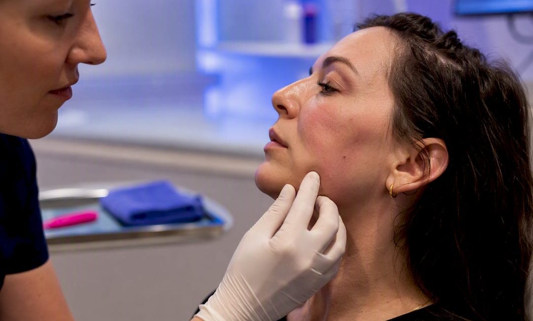
Most dermal filler reactions are mild and settle within days; the rare exceptions, vascular occlusion and infection, need urgent clinical attention. A systematic review of 3,967 patients found the overwhelming majority of reported events were simple injection-site reactions. If you or a patient notices worsening pain, skin colour change, or visual disturbance after treatment, contact the treating clinician or emergency services immediately. Delay is the single biggest driver of permanent harm in the small number of serious cases.
TL;DR:
- Most dermal filler complications are mild and resolve within two weeks, but vascular occlusion and infection require immediate intervention to prevent permanent damage.
- Vascular occlusion signs include severe pain, skin blanching, slow capillary refill, and expanding discoloration, necessitating urgent hyaluronidase and possibly ophthalmology referral.
- The highest risk zones for serious complications are the glabella, nose, lips, and temple due to limited collateral blood flow, especially when injecting near key arteries.
- Practitioners must have hyaluronidase readily available, follow strict protocols, and have emergency plans, as delayed treatment in vascular occlusion can lead to tissue necrosis or blindness.
- Clinic safety depends more on practitioner skill, proper training, and emergency readiness than on product choice alone.
Table of Contents
- Common dermal filler complications and how timing changes the picture
- Vascular occlusion: mechanisms, at-risk zones and the signs that separate it from bruising
- Infection, nodules and delayed hypersensitivity reactions
- How clinicians diagnose and treat filler complications
- Choosing a practitioner: questions to ask before you say yes
- What recovery actually looks like
- Clinic safety checklist for licensed practitioners
- Long-term complications: granulomas, chronic inflammation and migration
- Who is at higher risk, and why it matters
- Managing the rare but severe: blindness and tissue necrosis protocols
- Follow-up care after treatment
- Consent, documentation and the legal side of filler treatment
- Why safety literacy matters more than product choice
- Sources
- FAQ
Common dermal filler complications and how timing changes the picture
Complications are usually grouped by when they appear, and that timing tells you almost everything about how worried to be. Clinicians typically split them into three windows: immediate (within 24 hours), early (24 hours to 4 weeks), and delayed (beyond 4 weeks). Each window carries a different set of likely causes and a different urgency threshold.
Immediate reactions are overwhelmingly injection site reactions, or ISRs. These are the bruising, swelling, redness, and tenderness that almost everyone gets to some degree, plus the occasional Tyndall effect, a bluish discolouration that occurs when superficially placed hyaluronic acid filler scatters light under thin skin. Early complications can include delayed bruising, firmer swelling, or the first signs of an inflammatory nodule. Delayed complications, sometimes appearing months or even years later, include biofilm-related nodules, granulomas, and rarely, product migration.
The scale of the systematic review referenced above puts this in perspective. Across 38 studies covering nearly 4,000 patients, researchers recorded 8,795 complications, of which 7,719 were mild injection-site reactions and only 1,076 were classed as adverse events requiring some form of intervention. That is a ratio of roughly seven mild reactions for every one that needed active management.
Common injection site reactions include:
- Bruising, usually resolving within 7 to 14 days without treatment
- Swelling, often peaking at 24 to 48 hours post-treatment
- Erythema (redness) around the injection points
- Localised pain or tenderness, typically mild and short-lived
- Induration (firmness) that can persist for several weeks in denser filler areas
- The Tyndall effect, a visible blue-grey tint from superficial placement
Most of these need nothing more than time, cold compresses in the first 24 to 48 hours, and paracetamol if pain is bothersome. Arnica gel or tablets are commonly used, though the evidence for their effectiveness is mixed rather than conclusive. What you should not do is massage a fresh bruise aggressively or apply heat in the first day, since both can worsen swelling.
Key figure: Of 8,795 complications recorded across the pooled studies, 7,719 (roughly 88%) were mild injection-site reactions that resolved without specific treatment. The remaining 1,076 adverse events were where hyaluronidase featured as the most commonly used targeted treatment.
The line to watch is when a “normal” reaction stops behaving normally. Swelling that keeps building past 72 hours, pain that increases rather than fades, or skin that starts to look mottled or unusually pale, all warrant a call to the treating clinician rather than a wait-and-see approach. For licensed practitioners, this is also the point at which documenting the timeline (with photographs) matters most, both for clinical tracking and for later review if a complication does progress.
Vascular occlusion: mechanisms, at-risk zones and the signs that separate it from bruising
Vascular occlusion happens through one of three mechanisms: intravascular embolism, extravascular compression, or vascular spasm. Of the three, intravascular injection, where filler enters a vessel directly and blocks blood flow, has the strongest support in the literature as the primary driver of ischaemic events. Extravascular compression occurs when a large filler bolus presses on a nearby vessel from outside, cutting off flow without the product ever entering the bloodstream. Vascular spasm is the least common of the three and tends to resolve faster, though it can still cause a frightening early presentation.
Some occlusions declare themselves within minutes. Others creep in over hours, particularly with extravascular compression, where swelling builds slowly enough that the pattern can be mistaken for ordinary post-procedure oedema until the skin colour changes give it away.
Anatomical zones carry disproportionate risk. The nose, nasolabial folds, glabella, lips, and temple account for most reported severe vascular events, and the reason comes down to blood supply architecture. These areas rely on named arteries with limited collateral circulation, meaning there are few alternative routes for blood to reach the tissue if the main vessel is blocked. The glabella and nasal region connect, via the supratrochlear and dorsal nasal arteries, to the ophthalmic artery, which is why filler placed in these zones carries the rare but devastating risk of visual loss if product is injected retrograde into the vessel.
Recognising the difference between routine bruising and a vascular event comes down to a handful of clinical signs:
- Pain out of proportion to the procedure. Ordinary injection discomfort fades quickly; vascular pain often intensifies and feels disproportionately severe for the area treated.
- Blanching, a sudden whitening of the skin, followed by a mottled, dusky, or violaceous discolouration as the tissue becomes starved of oxygen.
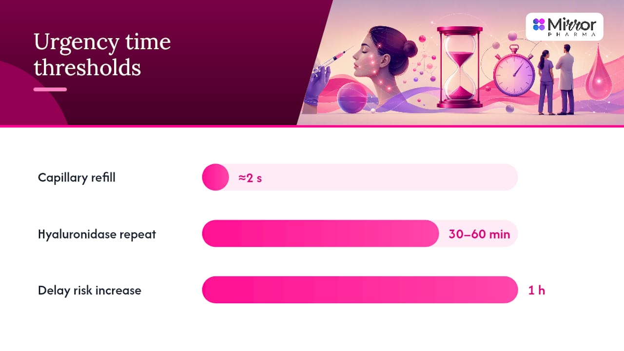
- Delayed capillary refill. Pressing the affected skin and releasing should see colour return within roughly one to two seconds; anything markedly slower points towards compromised flow, a distinction practitioner resources use as a quick bedside triage tool.
- An expanding area of discolouration, rather than one that stays static or fades, which suggests the compromised territory is growing.
- Coolness of the skin relative to the surrounding area, another sign the Cleveland Clinic’s patient guidance flags as worth immediate review.
Pro Tip: Compare the treated zone directly against an unaffected area of skin, side by side, rather than judging it in isolation. Vascular changes are often subtle at first, and a direct comparison makes blanching or duskiness far easier to spot early, when intervention still has the best chance of working.
Immediate steps for suspected occlusion follow a fairly consistent sequence across the literature. Stop injecting immediately. Mark the affected area with a pen so any spread can be tracked. Apply gentle massage and a warm compress to encourage vasodilation and flow. For hyaluronic acid fillers, hyaluronidase should be given urgently, and consensus guidance recommends high doses repeated at intervals rather than a single conservative dose. Low-dose aspirin is sometimes used as an adjunct to reduce clot propagation, though it does not replace hyaluronidase as the primary treatment. Any visual symptoms, blurring, pain behind the eye, or vision loss, mean an ophthalmology referral within minutes, not hours.
Incidence figures for vascular events are genuinely difficult to pin down. Published rates suggest they are rare, but survey data of experienced injectors indicates intravascular events happen more often than published figures capture, largely because many resolve with prompt treatment and are never formally reported. Treat any published “rare” figure as a floor, not a ceiling, when weighing your own risk tolerance around high-risk zones.
Infection, nodules and delayed hypersensitivity reactions
Not every lump under the skin after filler means infection. The two most common non-inflammatory causes are simple technique issues (filler placed too superficially, or unevenly) and over-correction, where too much product was used in one area. These lumps are usually soft, non-tender, and visible rather than painful, and often respond to gentle massage or, if persistent, a small dose of hyaluronidase for hyaluronic acid products.
Inflammatory nodules are a different problem. They present as firm, sometimes tender or warm lumps that can appear weeks to months after treatment, and they raise the question of biofilm formation, a thin layer of bacteria that adheres to the filler material and evades the immune system and standard antibiotics. Biofilms are notoriously difficult to diagnose because standard cultures often come back negative even when infection is present.
Working out which is which typically involves:
- Clinical examination comparing onset timing, texture, and tenderness against typical patterns
- Microbiology cultures, though these frequently underdetect biofilm-related organisms
- Imaging (usually ultrasound) if an abscess or fluid collection is suspected
- Extended-culture or specialist referral when atypical infection is suspected, since distinguishing inflammatory from infectious nodules can require tissue sampling and prolonged culture techniques, particularly with atypical mycobacteria
Treatment escalates with severity. Conservative nodules often respond to massage, time, or intralesional hyaluronidase for hyaluronic acid fillers. Confirmed or suspected infection needs antibiotics, chosen with input from microbiology where atypical organisms are suspected, since standard first-line agents can miss them entirely. Inflammatory (non-infectious) nodules sometimes respond to corticosteroids, though steroids should be used cautiously and only once infection has been reasonably excluded, since suppressing immune response in an infected nodule can worsen outcomes. A true abscess needs surgical drainage rather than antibiotics alone. Practitioners managing recurrent or atypical nodules should expect a longer treatment course, often stretching to weeks, and should set that expectation with the patient early rather than promising a quick resolution.
How clinicians diagnose and treat filler complications
Deciding how urgently to act starts at the bedside, not in a lab. The capillary refill test, pressing the skin and timing how quickly colour returns, remains the fastest way to flag possible vascular compromise, and a delay beyond roughly two seconds should prompt immediate escalation rather than a “wait and see” approach. Comparing the affected area to unaffected skin nearby sharpens the test further, since baseline refill time varies between individuals and body areas.
Point-of-care ultrasound has become an increasingly useful adjunct in this process. It lets a clinician confirm whether blood flow is actually present in a suspected occluded vessel and can help localise exactly where filler sits relative to the vessel, guiding more precise hyaluronidase placement rather than blind, widespread injection. It is not universally available in every clinic, but where it is, it meaningfully shortens the time between suspicion and confirmed diagnosis.
Treatment choice depends entirely on the diagnosis:
- Hyaluronidase for suspected vascular occlusion from hyaluronic acid filler, given urgently and often repeated at intervals until signs improve
- Antibiotics for confirmed or strongly suspected infection, adjusted based on culture results where available
- Corticosteroids or NSAIDs for inflammatory reactions once infection has been reasonably ruled out
- Surgical drainage for confirmed abscess formation
- Hyperbaric oxygen therapy as an adjunct in some severe ischaemic cases, though the evidence base for this remains limited to case reports rather than controlled trials
The phrase clinicians use internally is “time is tissue, time is vision.” Every hour of delay in a genuine vascular occlusion increases the risk of permanent skin necrosis, and for periorbital injections, increases the risk of irreversible vision loss. This is why expert consensus recommendations push so hard for clinics to have hyaluronidase on hand before a single injection is given, not ordered afterwards.
Pro Tip: If a suspected vascular event does not visibly improve within 30 to 60 minutes of the first hyaluronidase dose, repeat dosing is standard practice, not a sign the first dose failed. Waiting to “see if it works” for several hours before repeating treatment is one of the most common preventable delays in occlusion management.
Multi-disciplinary referral matters here too. Ophthalmology involvement for any visual symptom, plastic surgery or dermatology input for evolving necrosis, and infectious disease consultation for atypical or persistent infections all improve outcomes compared with a single practitioner managing a severe complication alone.
Choosing a practitioner: questions to ask before you say yes
The single biggest modifiable risk factor in filler complications is not the product, it is the person holding the needle. A 2026 analysis links irreversible complications predominantly to gaps in anatomical knowledge and incorrect injection technique, rather than to any particular filler brand or formulation. That puts the emphasis squarely on who you choose, not just what they use.
Before booking a treatment, or before a clinic books its next patient, these questions separate genuinely safety-conscious practice from a cosmetic add-on service:
- What are your qualifications, and are you a registered healthcare professional? Aesthetic injecting should be performed by someone with recognised medical, dental, or nursing training, not a beautician with a weekend certificate.
- Do you stock hyaluronidase on-site, and is it in date? A practitioner who cannot answer this immediately and confidently is not prepared for the rare event that it is needed.
- What is your emergency protocol if I show signs of vascular occlusion? A confident, specific answer (who to call, what steps happen first, where the nearest ophthalmology unit is) signals real preparedness.
- Do you use cannula or needle for this area, and why? Blunt cannulas reduce, but do not eliminate, the risk of intravascular injection in higher-risk zones compared with sharp needles, and a practitioner should be able to explain their technique choice for your specific treatment area.
- What is your follow-up policy? A clinic that books a check-in call or review appointment, rather than one that disappears after payment, is one that takes post-treatment monitoring seriously.
- Can I see your consent documentation and product information? You should be shown the product name, batch number, and given written information on risks before treatment, not just verbally told “it’s very safe.”
Red flags worth walking away from include unregulated or non-clinical premises, a practitioner who becomes defensive or vague when asked about emergency protocols, and any unwillingness to discuss consent documentation openly. Legitimate providers welcome these questions; hesitation is itself useful information.
Pre-procedure precautions that reduce risk on the patient side include reviewing current medications (particularly blood thinners, which increase bruising risk) with the treating clinician, and disclosing any active skin infection, since injecting through compromised skin raises infection risk substantially. Immediately post-procedure, avoiding strenuous exercise, alcohol, and excessive heat exposure (saunas, hot yoga) for 24 to 48 hours reduces swelling and bruising. For a deeper look at technique choice specifically, this comparison of cannula versus needle safety tradeoffs covers the clinical reasoning in more depth.
What recovery actually looks like
Ordinary injection site reactions resolve within one to two weeks in most patients, with bruising typically the longest-lasting element. Inflammatory nodules are less predictable. Some settle within a month with conservative management; others, particularly biofilm-related cases, can persist for months and require repeated treatment cycles before fully resolving.
Prognosis for vascular events is almost entirely a function of speed. Where hyaluronidase is given within the first few hours, most tissue makes a full recovery. Where treatment is delayed by many hours or longer, permanent skin necrosis and scarring become genuinely possible outcomes, and in the periorbital region, delayed treatment carries a real risk of permanent vision loss. This is not a complication where “it will probably sort itself out” is ever the right instinct.
Reconstructive or specialist input, dermatology for scarring, plastic surgery for tissue loss, becomes necessary in the minority of cases where necrosis has already progressed before treatment began. Early referral in these cases tends to produce better cosmetic and functional results than delayed specialist involvement.
Clinic safety checklist for licensed practitioners
For qualified aesthetic professionals, clinic-level preparedness is what actually reduces harm when a rare complication occurs. Consensus guidance is consistent on this: preparedness, not just product choice, is what changes outcomes.
A minimum emergency kit and protocol should include:
- In-date hyaluronidase stock, stored correctly and checked regularly
- A visible emergency protocol poster or laminated card at the treatment station
- Staff trained specifically to recognise early vascular signs, not just administer product
- Access to point-of-care ultrasound, or a clear referral pathway to one
- Up-to-date contact details for ophthalmology and plastic surgery referral
Mirror Pharma’s hyaluronic acid filler safety guide covers product selection and complication protocols in more detail for clinicians reviewing their own readiness. For clinics assessing product stock ahead of the next order cycle, the HyaCorp FACE dermal filler range is available to verified, licensed practitioners only.
Long-term complications: granulomas, chronic inflammation and migration
Some complications only show up long after the initial swelling has faded. Granulomas are firm nodules that form when the body mounts a chronic immune response to filler material, typically appearing months to years post-treatment rather than days. They differ from the biofilm nodules discussed earlier in that granulomas are a foreign-body immune reaction rather than an infection, though the two can be genuinely difficult to distinguish without tissue sampling.
Chronic low-grade inflammation, without a discrete lump, is another delayed pattern. It presents as persistent firmness or subtle swelling in the treated area that never fully settles, and it tends to respond slowly, if at all, to a single treatment. Management usually involves intralesional steroids, hyaluronidase for hyaluronic acid-based products, or a combination of both, with treatment sometimes needing to be repeated over several sessions.
Migration, where filler moves from its original placement site to a nearby area, is less common but well documented, particularly with higher-volume treatments or repeated touch-ups in the same region over years. It is usually a technique and product-selection issue rather than a sign of an acute complication, and correction typically involves dissolving the migrated product rather than attempting to reposition it. Patients with a history of multiple filler treatments in the same area over several years should be counselled that cumulative volume increases migration risk, a point worth discussing before repeat treatments rather than after migration has already occurred.
Who is at higher risk, and why it matters
Complication likelihood is not evenly distributed across every patient. Autoimmune conditions, including lupus and rheumatoid arthritis, are associated with a higher likelihood of inflammatory or granulomatous reactions to filler, since the immune system is already primed towards heightened responses to foreign material. Patients on immunosuppressive medication present the opposite concern: reduced ability to fight off infection if bacteria are introduced during injection, which raises the stakes around strict aseptic technique.
Previous filler or surgical procedures in the same area also raise risk, partly through scar tissue altering normal anatomy and vascular pathways, and partly through cumulative product volume increasing the chance of migration or granuloma formation discussed above. Patients with a history of cold sores (herpes simplex) around the lips are at increased risk of reactivation after lip filler specifically, and prophylactic antiviral medication is commonly considered for this group.
Smoking and poorly controlled diabetes both impair microvascular circulation, which matters enormously in a vascular occlusion scenario, since already-compromised blood flow gives less room for error if a vessel is partially obstructed. None of these factors make treatment impossible, but they change the risk conversation a licensed practitioner should have before proceeding, and they should be documented clearly in the patient’s pre-treatment assessment.
Managing the rare but severe: blindness and tissue necrosis protocols
Vision loss from filler is rare, but when it happens, it happens fast, usually within seconds to minutes of injection into a high-risk zone like the glabella or nose. The protocol on suspicion of visual involvement is immediate: stop injecting, call for emergency ophthalmology support without waiting to see if symptoms improve, and administer high-dose hyaluronidase around the affected vascular territory as fast as possible, even though its ability to reverse established retinal artery occlusion is limited once ischaemia has set in. Some protocols include retrobulbar hyaluronidase injection performed by trained ophthalmology staff, though this requires specific expertise and is not something a general aesthetic practitioner should attempt.
Tissue necrosis follows a slightly slower but still urgent timeline. The recommended approach is repeated, high-dose hyaluronidase injections at the affected site and surrounding territory, combined with warm compresses to encourage vasodilation, and close monitoring over the following 24 to 72 hours to see whether skin colour and capillary refill normalise. Photographic documentation at regular intervals helps track whether the compromised area is shrinking or expanding, information that directly guides whether treatment is working or needs escalation. Where skin breakdown has already begun, wound care and, in some cases, plastic surgery referral for debridement or reconstruction becomes part of the ongoing management plan.
Follow-up care after treatment
Most patients need nothing more than a check-in message or call within 48 to 72 hours to confirm swelling is improving rather than worsening. For higher-risk treatment areas, nose, glabella, or repeat treatments in previously treated zones, a same-day or next-day follow-up call is worth building into standard protocol rather than waiting for the patient to raise concerns themselves.
Patients should be given clear written guidance on what “normal” recovery looks like and, critically, a direct contact method for the treating clinician (not just a generic clinic reception line) if something feels wrong outside office hours. Vascular occlusion does not wait for opening hours, and a patient who cannot reach anyone quickly loses valuable time in exactly the scenario where minutes matter most.
For nodules or delayed reactions, a longer monitoring window applies, often with a review appointment at two to four weeks and again at three months if a nodule has not fully resolved. Clinics should document each follow-up interaction, including phone calls, since this record becomes part of the ongoing clinical picture if a complication does progress and needs escalated treatment later.
Consent, documentation and the legal side of filler treatment
Informed consent for filler treatment needs to go beyond a signature on a form. Patients should understand, in plain language, that dermal filler complications range from common and mild to rare and serious, and that no aesthetic treatment can be described as risk-free or guaranteed in outcome. Consent documentation should record the specific product used, including batch number, the treatment areas, and a summary of the risks discussed, including vascular occlusion and infection, however rare.
Photographic records before and after treatment support both clinical monitoring and, if needed, a clear timeline should a complication arise later. Practitioners should document any pre-existing conditions disclosed by the patient (autoimmune disease, previous filler, cold sore history) since these directly inform both the treatment plan and the consent conversation. Where a complication does occur, contemporaneous notes, what was seen, when, and what action was taken, matter enormously both for clinical continuity and for any subsequent review. This is standard practice for licensed practitioners and training academies alike, and it is worth treating documentation as part of patient safety rather than as an administrative afterthought.
Why safety literacy matters more than product choice
The conventional advice on filler safety tends to fixate on which product is “safest,” as if the molecule itself were the deciding factor. That framing misses what the evidence actually shows. The 2026 anatomical knowledge analysis points squarely at practitioner skill and technique, not product selection, as the dominant factor in irreversible harm. A well-chosen filler in inexperienced hands carries more risk than a well-known product handled by someone who understands facial vascular anatomy cold.
What gets underestimated is preparedness. Clinics obsess over technique training and skip the boring part, stocking hyaluronidase correctly, running emergency drills, having ophthalmology contacts on speed dial, because the rare event feels theoretical until it isn’t. The systematic review data showing that roughly seven in eight complications are mild should not breed complacency about the other one in eight. If you take one thing from this article, prioritise the clinic’s emergency readiness over the marketing claims on the product box.
— Rizwan
This article is general information, not a substitute for advice from a qualified doctor. Consult a qualified healthcare professional about your own circumstances before acting on anything here.
Sources
- Safety and potential complications of facial wrinkle correction with dermal fillers: a systematic literature review
- Treatment of Soft Tissue Filler Complications: Expert Consensus Recommendations
- Management of a vascular occlusion associated with cosmetic injections
- What is a vascular occlusion? (Cleveland Clinic patient information)
FAQ
What can go wrong with dermal fillers?
Most reactions are mild injection site effects like bruising, swelling, and tenderness that resolve within one to two weeks. Rarer complications include nodules, infection, and vascular occlusion, which can cause tissue damage or, very rarely, vision loss if not treated promptly.
Does filler ever fully leave your body?
Hyaluronic acid fillers are gradually broken down by the body over time, typically lasting several months to over a year depending on the product and area treated, though the exact rate varies between individuals. They can also be actively dissolved sooner using hyaluronidase if a complication or unwanted result occurs.
Are fillers more risky than anti-wrinkle injections?
The risk profiles differ rather than one being simply “more risky.” Filler carries a specific risk of vascular occlusion because it is injected as a physical volume that can block a vessel, a mechanism that does not apply to anti-wrinkle injections in the same way.
What is the riskiest place for fillers?
The glabella (between the eyebrows) and the nose carry the highest risk of serious vascular complications, including rare cases of vision loss, because they connect to the ophthalmic artery through limited collateral circulation. The lips, nasolabial folds, and temple are also considered higher-risk zones requiring careful technique.